
Acute Coronary Syndrome Pathway
From the first ECG to the discharge prescription: chest pain, STEMI, NSTE-ACS and the complications · v1- Say what the situation is first: undifferentiated chest pain, a STEMI on the ECG, a non-ST-elevation ACS, or a complication during the admission. The tool then asks only what that situation needs.
- Chest pain: it reads the ECG, runs the high-sensitivity troponin 0/1 h algorithm for the assay you name, and scores HEART.
- STEMI starts the clock. It chooses between primary PCI and fibrinolysis from the time to the nearest catheterisation laboratory, checks every contraindication, prints the weight-banded lytic dose with the co-therapy, and plans the transfer.
- Non-ST-elevation ACS: it computes GRACE and times the angiography.
- Name a complication and it names the treatment.
- Reading the ECG for you. It takes your reading as an entry, and the tracing itself decides everything downstream.
- Patients below 18 years, in whom chest pain is almost never coronary.
- Spontaneous coronary artery dissection, MINOCA and takotsubo syndrome. It names them where the entered picture raises them, and refers onward.
- The procedural detail of PCI and bypass surgery, the settings of mechanical circulatory support, and the management of chronic coronary syndromes after the first year.
1. The Situation and the Patient
1. The ECG: Patterns of Ischaemia and the STEMI Criteria
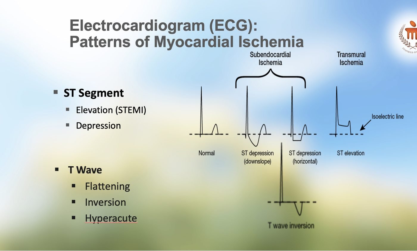
ST elevation is measured at the J point in two contiguous leads. The fourth universal definition sets the thresholds below; the other leads take 1 mm in every patient.
| Leads | Threshold | Note |
|---|---|---|
| V2 to V3 | 2 mm in men 40 and above; 2.5 mm in men under 40; 1.5 mm in women | The one lead pair with age and sex thresholds, because normal early repolarisation is largest here |
| All other leads | 1 mm | Two contiguous leads of the same territory |
| V7 to V9, posterior | 0.5 mm | Record them whenever V1 to V3 show ST depression with tall R waves |
| V3R to V4R, right-sided | 0.5 mm; 1 mm in men under 30 | Record them in every inferior infarct: the right ventricle changes the fluid and nitrate decisions |
| LBBB or paced rhythm | Sgarbossa: concordant ST elevation 1 mm or more (5 points); ST depression 1 mm or more in V1 to V3 (3 points); discordant ST elevation 5 mm or more (2 points). A score of 3 or more is specific | The modified rule replaces the 5 mm criterion with an ST to S-wave ratio of 0.25 or more, which is more sensitive |
| STEMI equivalents | de Winter: upsloping ST depression with tall symmetrical T waves, V1 to V6. Widespread ST depression with elevation in aVR | Both call for the STEMI pathway when the story fits |
2. High-Sensitivity Troponin: the ESC 0/1 h Algorithm
Rule-out at presentation needs a very low value and at least 3 hours since symptom onset. At 1 hour it needs a low 0 h value and a small change. Rule-in needs a high 0 h value or a large change. Everything between is the observe zone: a third sample at 3 hours, an echocardiogram and clinical judgement. Values in ng/L.
| Assay | Very low | Low | 1 h change, rule-out below | High | 1 h change, rule-in at or above |
|---|---|---|---|---|---|
| hs-cTnT, Roche Elecsys | below 5 | below 12 | 3 | 52 or more | 5 |
| hs-cTnI, Abbott Architect | below 4 | below 5 | 2 | 64 or more | 6 |
| hs-cTnI, Siemens Centaur | below 3 | below 6 | 3 | 120 or more | 12 |
| hs-cTnI, Beckman Access | below 4 | below 5 | 4 | 50 or more | 15 |
A conventional or point-of-care troponin cannot run this algorithm. A single negative result does not exclude infarction; the second sample belongs at least 6 hours after the first and at least 6 hours after the onset of pain.
3. The HEART Score
| Item | 0 | 1 | 2 |
|---|---|---|---|
| History | Slightly suspicious | Moderately suspicious | Highly suspicious |
| ECG | Normal | Non-specific repolarisation change, LBBB, LVH, digoxin | Significant ST deviation not explained by those |
| Age | Below 45 | 45 to 64 | 65 and above |
| Risk factors | None | One or two | Three or more, or known atherosclerotic disease |
| Troponin | At or below the reference limit | 1 to 3 times the limit | Above 3 times the limit |
0 to 3: major adverse cardiac events in about 1 to 2 per cent within 6 weeks in the validation cohort. 4 to 6: about 12 to 17 per cent. 7 to 10: about 50 to 65 per cent. The score was derived and validated in Dutch emergency departments; it has not been validated in an Indian cohort.
4. The GRACE In-Hospital Mortality Score
| Variable | Bands and points |
|---|---|
| Age, years | below 30: 0; 30 to 39: 8; 40 to 49: 25; 50 to 59: 41; 60 to 69: 58; 70 to 79: 75; 80 to 89: 91; 90 and above: 100 |
| Heart rate, per minute | below 50: 0; 50 to 69: 3; 70 to 89: 9; 90 to 109: 15; 110 to 149: 24; 150 to 199: 38; 200 and above: 46 |
| Systolic blood pressure, mmHg | below 80: 58; 80 to 99: 53; 100 to 119: 43; 120 to 139: 34; 140 to 159: 24; 160 to 199: 10; 200 and above: 0 |
| Creatinine, mg/dL | below 0.4: 1; 0.4 to 0.79: 4; 0.8 to 1.19: 7; 1.2 to 1.59: 10; 1.6 to 1.99: 13; 2 to 3.99: 21; 4 and above: 28 |
| Killip class | I: 0; II: 20; III: 39; IV: 59 |
| Cardiac arrest at admission | 39 |
| ST-segment deviation | 28 |
| Raised cardiac markers | 14 |
Non-ST-elevation ACS: low risk 108 or below (in-hospital mortality below 1 per cent), intermediate 109 to 140 (1 to 3 per cent), high above 140 (above 3 per cent). STEMI: low 125 or below, intermediate 126 to 154, high above 154. A GRACE score above 140 is the guideline threshold for angiography within 24 hours.
5. Reperfusion in STEMI: the Clock and the Choice
| Situation | Strategy | Target |
|---|---|---|
| Within 12 h of onset, PCI hospital | Primary PCI | Wire crossing within 60 min of diagnosis |
| Within 12 h, transfer can reach wire crossing within 120 min of diagnosis | Transfer for primary PCI | Wire crossing within 90 min of diagnosis; leave the referring hospital within 30 min |
| Within 12 h, PCI cannot be reached within 120 min | Fibrinolysis, then transfer to a PCI centre at once: the pharmacoinvasive strategy | Bolus within 10 min of diagnosis. Angiography 2 to 24 h after a successful lysis; rescue PCI at once when it fails |
| Cardiogenic shock or severe heart failure | Primary PCI whatever the delay. Lysis only where no PCI centre can be reached within 120 min and a mechanical complication is excluded | Immediate |
| 12 to 48 h after onset | Primary PCI if symptoms, instability or ischaemia continue; a routine primary PCI strategy should be considered even when settled. Fibrinolysis is not recommended | As soon as practicable |
| Beyond 48 h, settled, no ischaemia | No routine PCI of an occluded infarct artery; angiography on symptoms, viability or inducible ischaemia | Elective |
Failed fibrinolysis: less than 50 per cent ST resolution at 60 to 90 minutes in the lead with the greatest elevation, or persistent pain, or instability. That patient goes for rescue PCI immediately, which is why the transfer is arranged before the response is known.
6. Fibrinolytic Agents and Co-Therapy
| Agent | Dose | Notes |
|---|---|---|
| Tenecteplase | Single IV bolus over 5 to 10 seconds by weight: below 60 kg 30 mg; 60 to 69 kg 35 mg; 70 to 79 kg 40 mg; 80 to 89 kg 45 mg; 90 kg and above 50 mg. Half the dose at 75 years and above | Reconstituted at 5 mg/mL, so 30 mg is 6 mL. The half dose in the elderly came from the STREAM amendment after excess intracranial bleeding |
| Streptokinase | 1.5 million units IV over 30 to 60 minutes | The commonest lytic in India. Hypotension during the infusion: slow it, raise the legs, fluid. Never repeat in a patient who has had it before, at any time |
| Reteplase | 10 units IV bolus, repeated once after 30 minutes | Each bolus over 2 minutes |
| Alteplase, accelerated | 15 mg IV bolus, then 0.75 mg/kg over 30 minutes (maximum 50 mg), then 0.5 mg/kg over 60 minutes (maximum 35 mg); total not above 100 mg | The GUSTO regimen |
| Aspirin | 150 to 300 mg chewed, then 75 to 100 mg daily | The CSI writes 160 to 325 mg. A 325 mg tablet, or two of 150 mg, chewed |
| Clopidogrel | 300 mg loading, then 75 mg daily. At 75 years and above: no loading dose, 75 mg daily | Ticagrelor and prasugrel have no evidence with lysis and are not given with it; a switch may be considered 48 hours after lysis once PCI is done |
| Enoxaparin | Below 75 years: 30 mg IV bolus, then after 15 minutes 1 mg/kg SC twice daily, the first two doses capped at 100 mg. At 75 and above: no bolus, 0.75 mg/kg SC twice daily, the first two doses capped at 75 mg. Creatinine clearance below 30 mL/min: 1 mg/kg once daily | Until revascularisation or discharge, up to 8 days |
| Unfractionated heparin | 60 IU/kg IV bolus, maximum 4000 IU, then 12 IU/kg/h, maximum 1000 IU/h, to an aPTT of 1.5 to 2 times control for 24 to 48 hours | The alternative when enoxaparin is unsuitable |
| Fondaparinux | 2.5 mg IV bolus, then 2.5 mg SC once daily up to 8 days or discharge | Only with streptokinase, or no lytic; not with the fibrin-specific agents, and not in a creatinine clearance below 30 |
7. Antithrombotics with Primary PCI and in NSTE-ACS
| Drug | Dose | Where it sits |
|---|---|---|
| Aspirin | 150 to 300 mg chewed, then 75 to 100 mg daily | Every patient without a true allergy |
| Prasugrel | 60 mg loading, then 10 mg daily; 5 mg daily at 75 years and above or below 60 kg | Preferred with PCI. Contraindicated after any stroke or TIA. Given once the anatomy is known, not upstream |
| Ticagrelor | 180 mg loading, then 90 mg twice daily | Preferred with PCI; may be loaded before the anatomy is known in STEMI. Dyspnoea and bradycardia are the reasons it is stopped |
| Clopidogrel | 600 mg loading (300 mg with lysis), then 75 mg daily | When the potent agents are unavailable or contraindicated, with an oral anticoagulant, and with lysis |
| Unfractionated heparin | 70 to 100 IU/kg IV bolus at PCI; 50 to 70 IU/kg with a glycoprotein IIb/IIIa inhibitor | The standard anticoagulant for primary PCI and for NSTE-ACS going to the laboratory |
| Enoxaparin | 1 mg/kg SC twice daily; 0.5 mg/kg IV at PCI | NSTE-ACS awaiting angiography; renal adjustment as above |
| Fondaparinux | 2.5 mg SC once daily | NSTE-ACS where angiography is not early, with a UFH bolus at the time of PCI. Not in STEMI going for primary PCI |
Pretreatment: in NSTE-ACS whose anatomy is unknown and whose angiography is planned within 24 hours, routine P2Y12 loading before the laboratory is not recommended. Where the angiography will be delayed, loading may be considered. Dual antiplatelet therapy runs 12 months by default; at high bleeding risk it is shortened, or aspirin is dropped and a P2Y12 inhibitor continued alone from 1 to 3 months.
8. Secondary Prevention: the Discharge Prescription
| Measure | What | Target or duration |
|---|---|---|
| Antiplatelets | Aspirin 75 to 100 mg daily lifelong, plus a P2Y12 inhibitor | 12 months dual therapy by default |
| Statin | High intensity from the first day: atorvastatin 40 to 80 mg or rosuvastatin 20 to 40 mg; add ezetimibe 10 mg if the goal is not met at 4 to 8 weeks, then a PCSK9 inhibitor or inclisiran | LAI: LDL-C below 50 mg/dL. ESC: below 55 mg/dL and at least 50 per cent lower. ACC/AHA: add a non-statin at 70 mg/dL or above, consider from 55 |
| Beta-blocker | Metoprolol, bisoprolol or carvedilol once haemodynamically stable | Recommended with LVEF 40 per cent or below; with a preserved ejection fraction the benefit is uncertain since REDUCE-AMI, and the ESC keeps routine use as a weaker recommendation |
| ACE inhibitor or ARB | Ramipril, enalapril or an ARB, from the first 24 hours | Recommended with heart failure, LVEF 40 per cent or below, diabetes, hypertension or chronic kidney disease; reasonable for all |
| Mineralocorticoid antagonist | Eplerenone or spironolactone | LVEF 40 per cent or below with heart failure or diabetes |
| Blood pressure and glucose | Treat to target; an SGLT2 inhibitor or GLP-1 agonist where diabetes coexists | Below 130/80 mmHg; HbA1c below 7 per cent |
| Rehabilitation and lifestyle | Referral to cardiac rehabilitation before discharge; complete smoking cessation with pharmacotherapy; diet, activity, weight | Start within 2 weeks; 30 minutes of walking most days once cleared |
| Vaccination | Influenza vaccine during the admission or at the first review | Yearly |
9. Complications: the First Moves
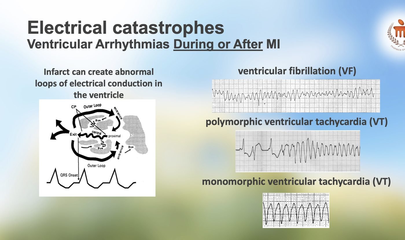
| Complication | First moves | Point to note |
|---|---|---|
| VF or pulseless VT | Unsynchronised shock at once; CPR between shocks; amiodarone 300 mg IV after the third shock, a further 150 mg if it recurs; correct potassium and magnesium; reperfuse | Early VF within 48 h does not by itself call for a defibrillator; late VF or VT does |
| Sustained monomorphic VT with a pulse | Unstable: synchronised cardioversion. Stable: amiodarone 150 mg IV over 10 minutes, then an infusion; a beta-blocker where the pressure allows; repeat cardioversion if it persists | Recurrent or incessant VT after the acute phase is a reason for urgent revascularisation and electrophysiology review |
| Polymorphic VT | Shock if pulseless; IV beta-blocker; potassium above 4 mmol/L and magnesium above 2 mg/dL; amiodarone; urgent angiography, because the substrate is ongoing ischaemia. With a long QT, magnesium 2 g IV and pace faster | Not a scar rhythm: treat the artery |
| Atrial fibrillation, fast | Unstable: cardioversion. Stable: a beta-blocker, or amiodarone where the ventricle is poor; anticoagulate on the CHA₂DS₂-VASc score | Treat the driver: heart failure, ischaemia, pericarditis |
| Bradycardia and AV block | Atropine 0.5 mg IV, repeated to 3 mg; transcutaneous pacing bridging to a temporary wire when symptomatic or high grade; reperfuse | Inferior: nodal, usually narrow, usually temporary. Anterior: infranodal, wide, a pacing indication, and a permanent device if the block has not resolved after 5 days |
| Cardiogenic shock | Immediate revascularisation of the culprit artery only; noradrenaline first, dobutamine for output; echocardiography now to exclude a mechanical cause; no routine balloon pump; a microaxial pump in selected STEMI shock at a centre that has one | SHOCK, CULPRIT-SHOCK, IABP-SHOCK II and DanGer Shock each changed one line of this row |
| Acute pulmonary oedema | Sit up, oxygen to 90 per cent or above, IV nitrate if systolic above 90, IV furosemide, non-invasive ventilation; no beta-blocker until dry; revascularise | Killip III: in-hospital mortality was 38 per cent in 1967 and is still several times that of class I |
| Right ventricular infarction | Fluid in 200 to 500 mL boluses to a systolic above 90; no nitrate, no diuretic, no morphine; pace for AV block; dobutamine if fluid fails; reperfuse | V4R in every inferior infarct |
| Mechanical complication | Echocardiography at the bedside, cardiac surgery called, vasodilator and balloon pump as a bridge if the pressure allows; pericardiocentesis is not the answer to a free wall rupture | Days 2 to 7; a new murmur, a sudden fall, or a tamponade |
| Pericarditis | High-dose aspirin, colchicine; avoid NSAIDs and corticosteroids in the first weeks | Early pericarditis in the first days; the delayed immune form at 2 to 8 weeks |
| LV thrombus | Warfarin or acenocoumarol for 3 to 6 months with repeat imaging; a DOAC may be considered | Anterior apical infarcts; the antiplatelet plan is shortened to fit |
1. One Umbrella, Three Diagnoses, Two Clocks
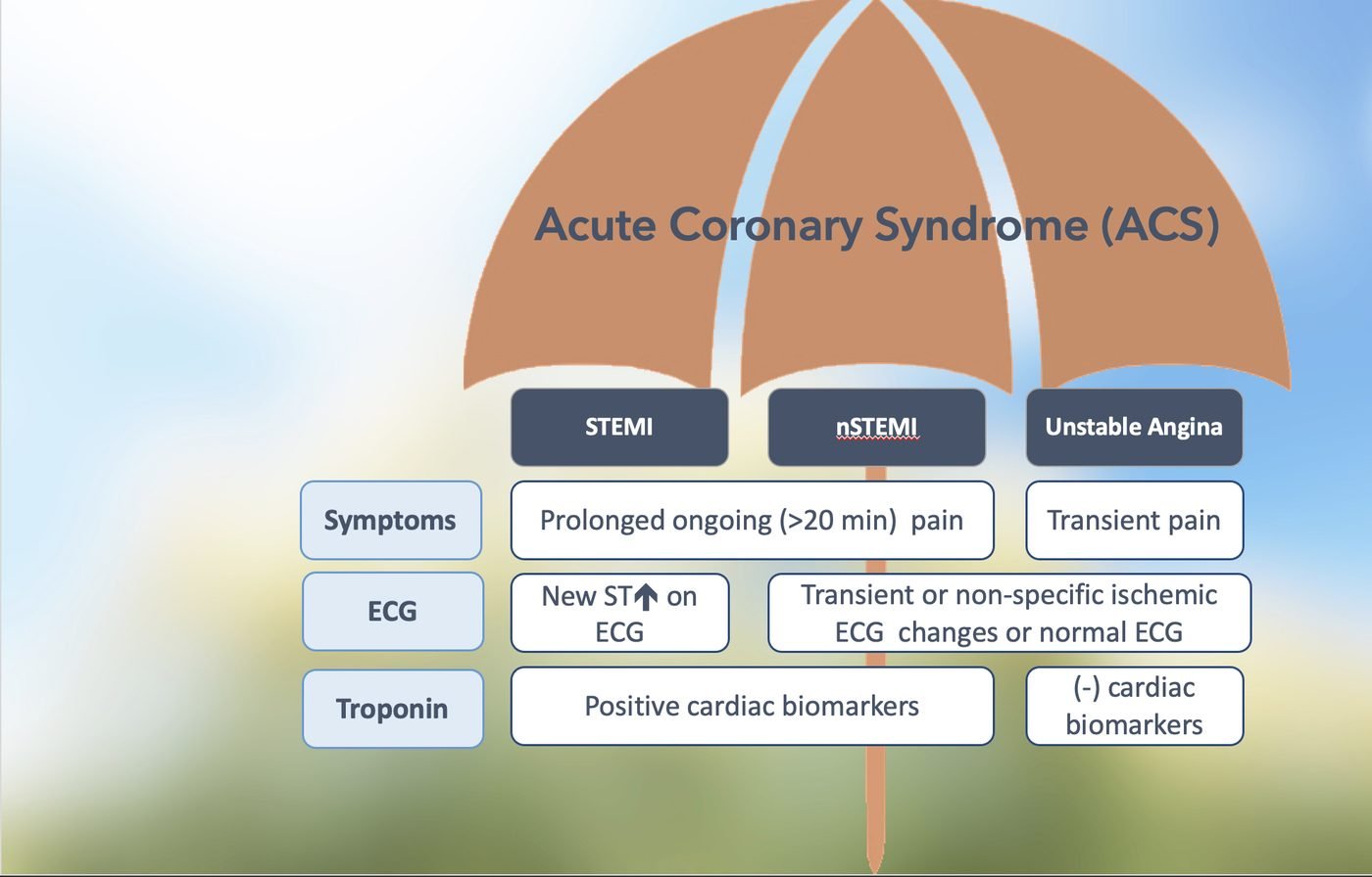
Plaque rupture or erosion with thrombus is one disease along a spectrum: the labels describe how much artery is blocked and for how long, not different illnesses. The ECG decides the first fork in minutes, persistent ST elevation meaning an occluded artery and a reperfusion clock; everything else waits for the troponin. The 2023 ESC guideline folded the STEMI and NSTE-ACS documents into one for that reason, and this tool follows. Four ECG patterns are occlusions that the voltage criteria send to the wrong lane.
| Pattern | What it means |
|---|---|
| Sgarbossa, GUSTO 1996 | The three features that show through a left bundle branch block: concordant ST elevation, ST depression in V1 to V3, grossly discordant elevation |
| Smith modification | Replaces the 5 mm absolute criterion with a ratio of ST elevation to S-wave depth of a quarter or more, raising sensitivity from about half to nine in ten |
| de Winter | Upsloping ST depression with tall T waves in the chest leads: a proximal LAD occlusion with no ST elevation at all |
| Wellens | Deep or biphasic T waves in V2 to V3 once the pain has settled: a critical LAD stenosis that will occlude if provoked. The laboratory, not a treadmill |
2. Time Is Muscle, and the Number Is 120
DeWood's angiograms in 1980 found a fresh occluding clot in nearly nine of ten transmural infarctions, fewer as the hours passed. The artery is shut and the clot is dissolving on its own, which is what puts a clock on the decision.
- 120 minutes is the exchange rate, from the diagnostic ECG to the wire crossing. Beyond it the advantage of primary PCI is spent and a lytic given within 10 minutes does better, particularly in the first 3 hours after onset when the clot is fresh and the two strategies are close to equivalent.
- STREAM tested exactly that in early presenters who could not reach PCI within an hour: tenecteplase then transfer matched primary PCI on death, shock, heart failure and reinfarction at 30 days, at the cost of more intracranial bleeding until the dose was halved at 75 years and above.
- The Indian corollary from CREATE. The median patient reached hospital 6 hours after the pain began. The delay a tool cannot fix is the one before the door.
3. Lyse and Send: the Indian Standard, and What the Contraindication List Is For
Most Indian patients with STEMI present to a hospital without a catheterisation laboratory and most receive streptokinase: 96 per cent of lytics in CREATE, 95 per cent in the Tamil Nadu STEMI programme. That programme showed the district hospital's job does not end at the bolus.
| Tamil Nadu STEMI programme, 35 spokes to 4 PCI hubs | Before | After |
|---|---|---|
| Angiography | 35 per cent | 61 per cent |
| PCI | 30 per cent | 47 per cent |
| One-year mortality | 17.6 per cent | 14.2 per cent |
Transfer after lysis for angiography within 24 hours, with rescue PCI when lysis failed. The transfer is arranged when the lytic is drawn up, not when the ST segments fail to fall, which is why this tool prints it on every lytic result.
Intracranial haemorrhage after lysis runs at about 1 per cent and is the complication that kills or disables.
- The absolute contraindications are the histories that predict it: previous intracranial bleed, recent ischaemic stroke, a lesion in the skull, recent major trauma or surgery, active bleeding, dissection.
- The relative ones weigh the size of the infarct against the bleed, in both directions. The small inferior infarct in a light, elderly, hypertensive patient is where the haemorrhage happens; the large anterior infarct two hours old with a four-hour transfer ahead is where withholding the drug is the harm.
- The tool refuses a dose across an absolute contraindication and prints the relative ones beside the dose, so the weighing stays visible.
4. Stratifying the Non-Elevation Patient: Troponin, Then the Scores
The older assays asked whether troponin was present. High-sensitivity assays measure it in nearly everyone, so the question became how much and whether it is moving.
- The ESC 0/1 h algorithm. A very low value 3 hours into the pain rules out infarction; a low value unchanged at 1 hour does the same; a high value or a large change rules it in. About a quarter of patients are left in an observe zone for a third sample and an echocardiogram.
- The cut-offs are assay-specific to the nanogram, which is why this tool asks which analyser the laboratory runs rather than printing one number.
- A raised troponin with no rise or fall is chronic myocardial injury, not infarction. A rise from sepsis, tachyarrhythmia, renal failure or pulmonary embolism is a type 2 infarction or an injury, and does not earn a stent.
| Score | Question it answers |
|---|---|
| HEART | Undifferentiated chest pain in the emergency department. Five bedside items; 3 or less carries under 2 per cent risk of a major event in 6 weeks. Outperformed TIMI and GRACE for that job in its validation. Use it to decide who goes home |
| GRACE | The patient already diagnosed with an ACS. Eight variables predicting in-hospital and 6-month death; 140 is the threshold for the laboratory within 24 hours. Use it to decide who goes tonight |
| TIMI | Seven yes-or-no items. The simplest and the least discriminating |
| Killip | Graded on what can be heard and seen: no failure, crackles or a third sound, frank pulmonary oedema, shock. Mortality in the original 250 patients, 1967, ran 6, 17, 38 and 81 per cent. Reperfusion has roughly halved every figure and left the gradient intact, and the class still carries 59 points in GRACE |
None of the first three has been validated in an Indian population, and the tool says so on the result.
5. Timing of Angiography in NSTE-ACS: Downgraded, Not Abandoned
TIMACS randomised 3,031 patients to angiography within 24 hours or after 36 and found no difference overall, but a clear benefit in the highest-risk third, GRACE above 140. Reading the later trials, the 2023 ESC guideline moved the early invasive strategy for high-risk NSTE-ACS from class I to class IIa: angiography within 24 hours should be considered, not must be done.
- Immediate angiography within 2 hours stays class I for the very-high-risk patient: shock, refractory pain, acute heart failure from ischaemia, a life-threatening arrhythmia, a mechanical complication, or recurrent dynamic ST changes.
- The practical Indian reading. The very-high-risk patient is transferred now, whatever the hour; the high-risk patient on the next available day; the rest can be worked up where they are.
6. Oxygen, Morphine, Nitrate and the Beta-Blocker
- Oxygen is a drug for hypoxaemia, not for chest pain. DETO2X randomised 6,629 patients with suspected infarction and a saturation of 90 per cent or above to oxygen or air and found no difference in death at a year. The threshold is 90 per cent.
- Morphine relieves pain and delays the absorption of the oral antiplatelet given beside it. Keep it for pain that nitrate has not settled.
- The nitrate has three stop signs: a systolic below 90, a right ventricular infarct whose output is preload, and a phosphodiesterase inhibitor in the previous day or two. The sublingual tablet given without a right-sided lead has produced more than one inferior-infarct collapse on Indian wards. It is also withheld in Killip class IV.
- The beta-blocker waits until the patient is out of Killip class II, because COMMIT's early intravenous metoprolol prevented reinfarction and fibrillation and caused cardiogenic shock in equal measure.
The long-term beta-blocker rests on trials from before reperfusion, when infarcts were large and ventricles poor. Both current positions are printed beside the drug rather than one being quietly picked.
- REDUCE-AMI, 2024. 5,020 patients with an infarct and an ejection fraction of 50 per cent or above, randomised to metoprolol or bisoprolol or none: no difference in death or reinfarction over 3.5 years.
- ESC 2023, written before that result: class I for an ejection fraction of 40 per cent or below, a weaker recommendation for everyone else.
7. The Second Antiplatelet, and Two Doses That Need Care in India
PLATO gave ticagrelor its place: against clopidogrel in 18,624 patients with ACS, fewer vascular deaths, infarctions and strokes at 12 months and lower all-cause death, at the cost of more non-procedural bleeding. Prasugrel earned the same standing in PCI-treated patients and edged ticagrelor head to head, which is why the ESC prefers it once the anatomy is known.
- Neither has evidence with a lytic, and neither belongs in a patient on warfarin or with a previous stroke. There, clopidogrel remains the drug.
- Prasugrel drops to 5 mg at 75 years or above, or under 60 kg, which describes many Indian elders.
- Ticagrelor's breathlessness is mistaken for heart failure often enough to be worth naming at discharge.
8. Shock: Open the Artery, Only That Artery
| Trial | What it settled |
|---|---|
| SHOCK, 1999 | Emergency revascularisation, rather than stabilisation first, lowered mortality at 6 months |
| IABP-SHOCK II, 2012 | Retired the balloon pump, until then a class I device, by finding no effect on 30-day death at all |
| CULPRIT-SHOCK, 2017 | Opening the non-culprit vessels in the same sitting killed more patients than opening the culprit alone |
| DanGer Shock, 2024 | A microaxial flow pump lowered 180-day death in STEMI shock, 58.5 to 45.8 per cent, with a four-fold rise in severe bleeding, limb ischaemia and haemolysis |
The order of moves that follows: noradrenaline to a pressure, the culprit artery, an echocardiogram to find the mechanical cause you cannot afford to miss, and a device only in the centre that has one and the patient who fits the trial.
9. The Lipid Target Is Lower in India, on Purpose
Indian patients infarct a decade younger than Europeans, at cholesterol levels a Western table would call modest.
| Source | Secondary prevention LDL cholesterol goal |
|---|---|
| Lipid Association of India, 2020 | Below 50 mg/dL, and 30 mg/dL for the patient who infarcts again within a year despite it |
| ESC | Below 55 mg/dL and at least a 50 per cent fall |
| ACC/AHA | Add a second drug at 70 mg/dL, and consider it from 55 |
All three agree on the sequence: a high-intensity statin on day one, ezetimibe at the 4 to 8 week check, then a PCSK9 inhibitor or inclisiran, with a fasting panel after each change. The number most often missing from an Indian discharge summary is the follow-up lipid test, and it is the only way to know whether any of this was reached.
10. What This Tool Deliberately Does Not Do
- It does not interpret the ECG. Your reading goes in, and if it is wrong the plan is wrong, which is why the criteria are printed beside the entry.
- It does not run a troponin algorithm for an assay it does not know, and does not invent a reference limit for the HEART score.
- It does not compute the GRACE score with a creatinine it has not been given.
- It does not print a lytic dose after 12 hours, across an absolute contraindication, or for an agent that is not in the hospital.
- It names spontaneous coronary dissection, MINOCA and takotsubo where the picture raises them, and stops there.
- Where the guidelines disagree, the ESC, the ACC/AHA and the Indian position are printed side by side and the one followed is named.
Abbreviations
ACC (American College of Cardiology) · ACE (Angiotensin-Converting Enzyme) · ACS (Acute Coronary Syndrome) · AHA (American Heart Association) · aPTT (Activated Partial Thromboplastin Time) · ARB (Angiotensin Receptor Blocker) · AV (Atrioventricular) · CHA₂DS₂-VASc (Congestive Heart Failure, Hypertension, Age, Diabetes, Stroke, Vascular Disease, Age, Sex Category Score) · CPR (Cardiopulmonary Resuscitation) · CSI (Cardiological Society of India) · DOAC (Direct Oral Anticoagulant) · ECG (Electrocardiogram) · eGFR (Estimated Glomerular Filtration Rate) · ESC (European Society of Cardiology) · GLP-1 (Glucagon-Like Peptide-1) · GRACE (Global Registry of Acute Coronary Events) · HbA1c (Glycated Haemoglobin) · HEART (History, ECG, Age, Risk Factors, Troponin Score) · hs-cTn (High-Sensitivity Cardiac Troponin) · hs-cTnI (High-Sensitivity Cardiac Troponin I) · hs-cTnT (High-Sensitivity Cardiac Troponin T) · IABP (Intra-Aortic Balloon Pump) · IU (International Units) · IV (Intravenous) · JVP (Jugular Venous Pressure) · LAD (Left Anterior Descending Artery) · LAI (Lipid Association of India) · LBBB (Left Bundle Branch Block) · LDL-C (Low-Density Lipoprotein Cholesterol) · LV (Left Ventricular) · LVEF (Left Ventricular Ejection Fraction) · LVH (Left Ventricular Hypertrophy) · MI (Myocardial Infarction) · MINOCA (Myocardial Infarction with Non-Obstructive Coronary Arteries) · NSAID (Non-Steroidal Anti-Inflammatory Drug) · NSTE-ACS (Non-ST-Elevation Acute Coronary Syndrome) · NSTEMI (Non-ST-Elevation Myocardial Infarction) · P2Y12 (Platelet Adenosine Diphosphate Receptor Subtype) · PCI (Percutaneous Coronary Intervention) · PCSK9 (Proprotein Convertase Subtilisin/Kexin Type 9) · SC (Subcutaneous) · SGLT2 (Sodium-Glucose Co-Transporter 2) · SpO₂ (Oxygen Saturation by Pulse Oximetry) · STEMI (ST-Elevation Myocardial Infarction) · TIA (Transient Ischaemic Attack) · TIMI (Thrombolysis in Myocardial Infarction Score) · UFH (Unfractionated Heparin) · VF (Ventricular Fibrillation) · VT (Ventricular Tachycardia)References
- Byrne RA, Rossello X, Coughlan JJ, et al. 2023 ESC Guidelines for the management of acute coronary syndromes. Eur Heart J. 2023;44(38):3720-3826.
- Rao SV, O'Donoghue ML, Ruel M, et al. 2025 ACC/AHA/ACEP/NAEMSP/SCAI Guideline for the Management of Patients With Acute Coronary Syndromes. Circulation. 2025;151(13):e771-e862.
- Guha S, Sethi R, Ray S, et al. Cardiological Society of India: position statement for the management of ST elevation myocardial infarction in India. Indian Heart J. 2017;69 Suppl 1:S63-S97.
- Thygesen K, Alpert JS, Jaffe AS, et al. Fourth universal definition of myocardial infarction (2018). Eur Heart J. 2019;40(3):237-269.
- Xavier D, Pais P, Devereaux PJ, et al. Treatment and outcomes of acute coronary syndromes in India (CREATE): a prospective analysis of registry data. Lancet. 2008;371(9622):1435-1442.
- Alexander T, Mullasari AS, Joseph G, et al. A system of care for patients with ST-segment elevation myocardial infarction in India: the Tamil Nadu-ST-segment elevation myocardial infarction program. JAMA Cardiol. 2017;2(5):498-505.
- Mohanan PP, Mathew R, Harikrishnan S, et al. Presentation, management, and outcomes of 25 748 acute coronary syndrome admissions in Kerala, India: results from the Kerala ACS Registry. Eur Heart J. 2013;34(2):121-129.
- Puri R, Mehta V, Duell PB, et al. Proposed low-density lipoprotein cholesterol goals for secondary prevention and familial hypercholesterolemia in India with focus on PCSK9 inhibitor monoclonal antibodies: expert consensus statement from Lipid Association of India. J Clin Lipidol. 2020;14(2):e1-e13.
- ISIS-2 (Second International Study of Infarct Survival) Collaborative Group. Randomised trial of intravenous streptokinase, oral aspirin, both, or neither among 17,187 cases of suspected acute myocardial infarction: ISIS-2. Lancet. 1988;2(8607):349-360.
- The GUSTO Investigators. An international randomized trial comparing four thrombolytic strategies for acute myocardial infarction. N Engl J Med. 1993;329(10):673-682.
- Armstrong PW, Gershlick AH, Goldstein P, et al. Fibrinolysis or primary PCI in ST-segment elevation myocardial infarction. N Engl J Med. 2013;368(15):1379-1387.
- Killip T 3rd, Kimball JT. Treatment of myocardial infarction in a coronary care unit. A two year experience with 250 patients. Am J Cardiol. 1967;20(4):457-464.
- Granger CB, Goldberg RJ, Dabbous O, et al. Predictors of hospital mortality in the global registry of acute coronary events. Arch Intern Med. 2003;163(19):2345-2353.
- Backus BE, Six AJ, Kelder JC, et al. A prospective validation of the HEART score for chest pain patients at the emergency department. Int J Cardiol. 2013;168(3):2153-2158.
- Sgarbossa EB, Pinski SL, Barbagelata A, et al. Electrocardiographic diagnosis of evolving acute myocardial infarction in the presence of left bundle-branch block. N Engl J Med. 1996;334(8):481-487.
- Mehta SR, Granger CB, Boden WE, et al. Early versus delayed invasive intervention in acute coronary syndromes. N Engl J Med. 2009;360(21):2165-2175.
- Wallentin L, Becker RC, Budaj A, et al. Ticagrelor versus clopidogrel in patients with acute coronary syndromes. N Engl J Med. 2009;361(11):1045-1057.
- Hofmann R, James SK, Jernberg T, et al. Oxygen therapy in suspected acute myocardial infarction. N Engl J Med. 2017;377(13):1240-1249.
- Thiele H, Akin I, Sandri M, et al. PCI strategies in patients with acute myocardial infarction and cardiogenic shock. N Engl J Med. 2017;377(25):2419-2432.
- Møller JE, Engstrøm T, Jensen LO, et al. Microaxial flow pump or standard care in infarct-related cardiogenic shock. N Engl J Med. 2024;390(15):1382-1393.
- Yndigegn T, Lindahl B, Mars K, et al. Beta-blockers after myocardial infarction and preserved ejection fraction. N Engl J Med. 2024;390(15):1372-1381.
How to Cite This Tool
AMA Style:Umakanth S. Acute Coronary Syndrome Pathway. Version 1. MEDiscuss Clinical Decision Support System. Published 2026. Accessed . https://mediscuss.org/cdss/acs-pathway
Vancouver Style:Umakanth S. Acute Coronary Syndrome Pathway [Internet]. Version 1. MEDiscuss.org; 2026 [cited ]. Available from: https://mediscuss.org/cdss/acs-pathway
Save this calculation
Whatever you type here stays on this device and is not sent anywhere. The server receives only a scrambled code made from it, so nobody with access to the server can tell which patient a saved calculation belongs to. Enter the same nickname the next time to see that patient's earlier values. What is stored