
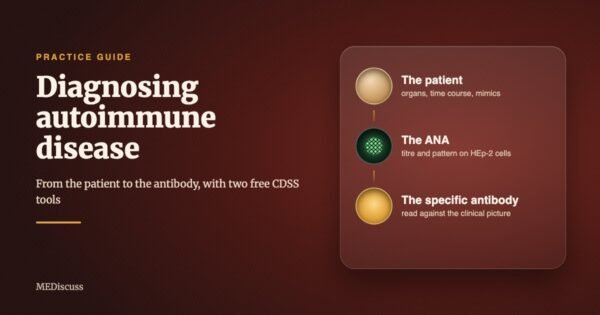
A young woman comes to the clinic with joint pains and a report that says ANA positive, 1:80. Does she have lupus? Most of the time, no. A man with fever, blood in the urine and a positive MPO-ANCA looks like vasculitis. Sometimes he has infective endocarditis instead, and steroids would harm him. Autoimmune disease is diagnosed from the patient first and from the antibody second, and most of the errors in this area come from reversing that order.
This guide is for medical students, interns and residents. It explains, in plain language, how autoimmune disease is diagnosed, what each common antibody test means, and where the traps lie in Indian practice. It then shows, step by step and with screenshots, how to use two free tools on the MEDiscuss clinical decision support system (CDSS) that were built for this job:
- Autoimmune Disease Workup Pathway: starts from the patient’s clinical picture and works through the ANA, the specific antibodies, the classification criteria and the next tests.
- Autoantibody Interpretation Guide: a simpler companion. You enter the antibodies a report shows as positive, and it tells you what each one, and each combination, usually means.
Both are free and need no sign-in. At the end there are nine worked cases. Each is a link that opens the tool with the case already filled in, so you can change one finding at a time and watch the reasoning move.
What autoimmunity means
The immune system is trained to attack what is foreign and to leave the body’s own tissues alone. That restraint is called tolerance. When tolerance fails, the immune system makes antibodies and T cells against the body’s own proteins. These are autoantibodies, and the illness that follows is an autoimmune disease.
Two points make the rest of this guide easier to follow.
- Autoimmune diseases come in two broad kinds. Some are systemic: lupus (SLE), Sjögren disease, systemic sclerosis, myositis, the ANCA vasculitides and antiphospholipid syndrome can involve skin, joints, kidneys, lungs, blood and brain in the same patient. Others are organ-specific: autoimmune hepatitis, primary biliary cholangitis, membranous nephropathy, pemphigus and pemphigoid, Guillain-Barré syndrome and autoimmune encephalitis mainly involve one organ.
- An autoantibody is not a disease. Many healthy people carry autoantibodies all their lives and never fall ill. Infections, drugs and cancers can also produce them. The antibody is a clue whose weight depends on who is carrying it.
Why the antibody is not the diagnosis
The most important idea in this whole field is pre-test probability: how likely the disease is before the test result is known. It decides what a positive result means, and nothing about the laboratory changes it.
Consider the antinuclear antibody (ANA). In a large survey of the United States population, 13.8 per cent of people aged 12 and over had a positive ANA at a 1:80 dilution. It was commoner in women (17.8 per cent) than in men (9.6 per cent) and rose with age [1]. Fifteen reference laboratories that tested healthy people found this [2]:
| Titre | Healthy people positive | What it means |
|---|---|---|
| 1:40 | 31.7 per cent | Almost one in three well people. Close to useless on its own. |
| 1:80 | 13.3 per cent | About one in eight well people. The entry point for the 2019 lupus criteria, not proof of disease. |
| 1:160 | 5.0 per cent | About one in twenty well people. |
| 1:320 | 3.3 per cent | Uncommon in health, but 3.3 per cent of a large population is still a lot of people. |
Now picture a clinic that tests 1,000 people, of whom 20 have lupus. With a sensitivity of 98 per cent and a specificity of 90 per cent, about 20 of the 20 with lupus test positive, and about 98 of the 980 without lupus also test positive. Of 118 positive results, only 20 are true. In that clinic, a positive ANA is wrong about five times out of six.
So before you order an ANA, ask a simple question: what will I do differently if it is positive, and what will I do differently if it is negative? If the answer to both is nothing, the test will not help that patient.
The ANA earns its place in the other direction. At 1:80 it is positive in about 98 per cent of people with lupus [3], so a negative ANA makes lupus very unlikely. It is a good test for ruling lupus out and a weak test for ruling it in.
The order of thinking
The figure below is the sequence both tools follow. The antibody is the third step, not the first.
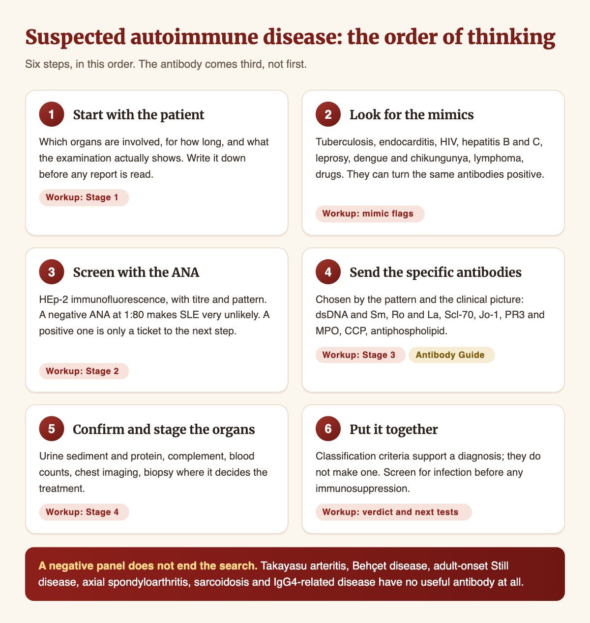
Step 1. Start with the patient
Write down which organs are involved and for how long, before you read any report. Ask about Raynaud phenomenon, rashes and photosensitivity, mouth ulcers, hair loss, dry eyes and mouth, joint swelling (not only pain), muscle weakness, breathlessness and cough, frothy or red urine, miscarriages and clots, and every drug the patient takes. Examine the joints, skin, nails and nail folds, lungs and pulses, and test the urine. A dipstick costs very little and finds nephritis early.
Step 2. Look for the mimics
In India, infections copy autoimmune disease often, and some of them turn the same antibodies positive. Steroids given for a presumed autoimmune illness that was really an infection are the commonest serious error in this area. The main mimics are listed later in this guide.
Step 3. Screen with the ANA
The ANA is a screening test for lupus and related diseases. It is covered in detail in the next section.
Step 4. Send the specific antibodies
Choose them from the ANA pattern and the clinical picture, not as a large panel for everyone. A patient with Raynaud phenomenon and tight skin needs a different panel from a patient with haemoptysis and red cells in the urine.
Step 5. Confirm and stage the organs
Urine microscopy and the protein-creatinine ratio, complement (C3 and C4), blood counts, liver tests, a chest X-ray or CT, an echocardiogram where the lungs or heart may be involved, and a biopsy where it will decide treatment.
Step 6. Put it together
Match the clinical picture, the antibodies and the organ findings. Use the classification criteria as a check, not a verdict, and screen for infection before any immunosuppression.
The ANA: three results in one report
A good ANA report gives you three things: the method, the titre and the pattern.
- Method. The reference method is indirect immunofluorescence on HEp-2 cells. Some laboratories screen with an ELISA or a chemiluminescence assay (CLIA) instead. These give a positive or negative result and no pattern.
- Titre. The highest dilution at which staining is still seen. A higher titre means more antibody, and it is less common in healthy people (see the table above).
- Pattern. What the staining looks like. It tells you what the antibody is directed against, and so which specific antibodies to send next. A titre reported without a pattern throws away half of the information the test produced.
The International Consensus on ANA Patterns (ICAP) gives each pattern an AC code [4]. The common ones:
- Homogeneous (AC-1): dsDNA, nucleosome, histone. Think of SLE, drug-induced lupus.
- Dense fine speckled (AC-2): DFS70, often alone. Usually a healthy person when anti-DFS70 is the only antibody.
- Centromere (AC-3): CENP-A, CENP-B. Think of limited cutaneous systemic sclerosis, primary biliary cholangitis.
- Speckled, fine or coarse (AC-4, AC-5): Ro, La, Sm, U1-RNP. Think of SLE, Sjögren disease, mixed connective tissue disease.
- Nucleolar (AC-8 to AC-10): Scl-70, PM-Scl, fibrillarin, Th/To. Think of systemic sclerosis and overlap.
- Nuclear dots or rim (AC-6, AC-7, AC-11, AC-12): sp100, gp210. Think of primary biliary cholangitis.
- Cytoplasmic (AC-15 to AC-23): Jo-1, ribosomal P, mitochondria, SRP. Think of myositis, primary biliary cholangitis.
Two traps are worth remembering.
- A cytoplasmic pattern may be reported as “ANA negative”, because strictly it is not nuclear. In a patient with myositis or unexplained interstitial lung disease, that “negative” report may be hiding an anti-Jo-1. Read the comment on the report, not only the conclusion [4].
- A dense fine speckled pattern is the commonest high-titre pattern in healthy people. If anti-DFS70 is confirmed and no disease-specific antibody is present, it argues against a systemic rheumatic disease. Isolated anti-DFS70 is found in 2 to 22 per cent of healthy people but in under 1 per cent of ANA-associated rheumatic disease [5].
One more point. The ANA does not track disease activity. Once it is positive, repeating it answers no question and costs the patient money.
The specific antibodies, and what each one tells you
Specific antibodies are what turn a vague picture into a named disease. The list below gives the ones you will meet most often. The figures come from the sources behind the two tools, where each is cited in full.
- Anti-dsDNA: SLE, lupus nephritis. Counts in the 2019 criteria only on an assay of documented 90 per cent specificity or better. Its level rises with nephritis flares, so it is worth repeating.
- Anti-Sm: SLE, almost only SLE. The most specific lupus antibody, but found in a minority of patients. Establishes the diagnosis; no need to repeat it.
- Anti-Ro60 (SSA) and anti-La (SSB): Sjögren disease, SLE, subacute cutaneous lupus. Anti-Ro in a woman who is or may become pregnant carries a risk of congenital heart block in the baby. Anti-La alone carries little weight.
- Anti-Ro52: Myositis, systemic sclerosis, interstitial lung disease. Not a Sjögren marker on its own. Look at the lungs.
- Anti-U1-RNP: Mixed connective tissue disease, SLE. High titre with no lupus-specific antibody suggests mixed connective tissue disease. Check the heart and pulmonary pressures.
- Anti-Scl-70, anticentromere, anti-RNA polymerase III: Systemic sclerosis. Scl-70: lung fibrosis. Anticentromere: pulmonary hypertension, later. RNA polymerase III: renal crisis and a cancer search.
- Anti-Jo-1 and other antisynthetase antibodies: Antisynthetase syndrome. Lung disease often dominates and can come before the muscle weakness. Get an HRCT whatever the CK.
- Anti-MDA5: Dermatomyositis with little muscle disease. Rapidly progressive lung disease with a near-normal CK. Treat it as a respiratory emergency.
- RF and anti-CCP: Rheumatoid arthritis. Anti-CCP is the more specific of the two. RF is positive in many infections and in hepatitis C.
- PR3-ANCA and MPO-ANCA: ANCA vasculitis. Ask for the antigen-specific immunoassay, not only a c-ANCA or p-ANCA pattern [6]. MPO-ANCA alone is the least specific result in the panel.
- Anti-GBM: Anti-glomerular basement membrane disease. An emergency. About a third are also ANCA positive.
- Lupus anticoagulant, anticardiolipin, anti-beta2-glycoprotein I: Antiphospholipid syndrome. Must be positive twice, at least 12 weeks apart [7]. All three positive is the highest-risk profile.
A useful habit is to sort antibodies into two groups. Static markers, such as the ANA, anti-Sm, anti-Ro, anti-Scl-70, the myositis antibodies and anti-CCP, establish the diagnosis and do not need repeating. Dynamic markers, such as the anti-dsDNA level, C3 and C4, the urine protein and sediment, and the CK in myositis, move with disease activity and are worth following.
A note on line blots. Most Indian laboratories report the ANA profile, the myositis panel and the neuronal panel as line blots, graded from + to ++++. A weak band (+ or ++) and a strong band (+++ or ++++) are not the same finding. Weak bands cross-react and are the commonest source of false positives. Record what the report actually says.
The mimics: rule them out before you name the disease
These conditions can look like autoimmune disease, and several of them turn autoantibodies positive. Each one has its own test that separates it.
- Tuberculosis. Chronic fever, weight loss, serositis, a cavitating lung lesion that looks like GPA, reactive arthritis. Can produce RF, a low-titre ANA and occasionally ANCA. What separates it: Tissue diagnosis, CBNAAT, culture. A cavity in India is tuberculosis until proven otherwise.
- Infective endocarditis. Fever, glomerulonephritis, low complement and a positive ANCA together. ANCA was found in 18 per cent in one series [8]. What separates it: Blood cultures before antibiotics, and echocardiography.
- Leprosy. Polyarthritis, neuropathy, and an erythema nodosum reaction that looks like vasculitis. False-positive antiphospholipid antibodies. What separates it: Slit-skin smear, thickened nerves, the pattern of the skin lesions.
- Dengue and chikungunya. Severe symmetric polyarthritis, rash, low platelets. Chikungunya arthritis can last months and is mistaken for early rheumatoid arthritis. What separates it: The epidemic setting, the acute febrile start, and serology for the virus.
- HIV, hepatitis B and hepatitis C. Cytopenias, arthritis, sicca, vasculitic rash. Hepatitis C often gives a strongly positive RF. What separates it: Serology, and HIV testing before immunosuppression without exception.
- Kikuchi-Fujimoto disease. A young woman with fever, tender neck nodes and low counts, closely copying a lupus flare. What separates it: Lymph node biopsy.
- Drugs. Hydralazine, isoniazid, procainamide, minocycline and anti-TNF agents can cause drug-induced lupus. Propylthiouracil and hydralazine can cause ANCA vasculitis. What separates it: A full drug history. Anti-histone antibodies are common in drug-induced lupus but are not specific for it.
Classification criteria are not diagnostic criteria
You will hear about the 2019 EULAR/ACR lupus criteria [9], the 2010 rheumatoid arthritis criteria [10], the 2016 Sjögren criteria [11] and the 2022 vasculitis criteria [12]. These were written to select clean groups of patients for research, so they favour specificity. A few rules make them easier to use:
- Most have an entry rule. The 2019 lupus criteria apply only if the ANA is 1:80 or higher on HEp-2 cells (or an equivalent positive test). The 2022 vasculitis criteria apply only once small- or medium-vessel vasculitis is already the working diagnosis.
- Items count only if nothing else explains them better. A fever from tuberculosis does not score for lupus.
- In the lupus criteria, only the highest item in each domain counts, and at least one clinical item is needed besides the 10 points.
- Meeting the criteria supports a diagnosis; missing them does not exclude one. The authors of the 2019 lupus criteria say the criteria should never be used to deny appropriate treatment to a patient who does not fully meet them.
When the antibody panel is negative
A negative panel does not mean “not autoimmune”. Several important diseases have no useful antibody at all:
- Takayasu arteritis: a young woman with limb claudication, weak or absent pulses, a blood pressure difference between the arms, or unexplained hypertension. Diagnosed by CT or MR angiography.
- Behçet disease: recurrent mouth and genital ulcers, uveitis, pathergy.
- Axial spondyloarthritis: inflammatory back pain starting before 45, with sacroiliac imaging and HLA-B27.
- Adult-onset Still disease: daily spiking fever, a salmon-pink evanescent rash, sore throat, arthritis and a high neutrophil count. A diagnosis of exclusion [13].
- Polymyalgia rheumatica and giant cell arteritis: an older patient with girdle stiffness, new headache or jaw claudication, and a high ESR or CRP.
- Sarcoidosis and IgG4-related disease: diagnosed on histology.
- ANCA-negative vasculitis and ANA-negative lupus: uncommon, but real.
Before any immunosuppression
The 2022 EULAR recommendations advise screening for hepatitis B in every patient, and for tuberculosis before a biologic, with tuberculosis screening to be considered before glucocorticoids and other immunosuppressants. Hepatitis C and HIV serology should be considered [14]. In Indian practice, tuberculosis screening before high-dose steroids is a sensible default. Add a pregnancy test before drugs such as mycophenolate, methotrexate or cyclophosphamide. The workup tool adds this screen to its list of next investigations automatically.
The two tools: which one to open
Autoimmune Disease Workup Pathway
- Start from the patient: age, sex and clinical features, then the tests.
- You enter clinical features in 12 groups, the ANA, the antibody panel and the confirmatory tests.
- You get a verdict, the classification criteria with each point shown, red flags, signposts for seronegative and organ-specific disease, and a list of next investigations.
- Probability: a post-test probability of SLE, shown only when the picture is lupus-like.
- Best for a new patient with a suspected systemic autoimmune disease.
- Short link: mediscuss.org/go/autoimmune
Autoantibody Interpretation Guide
- Start from an antibody report.
- You enter the ANA titre and pattern, and the antibodies reported positive (79 in all).
- You get the disease pattern the antibodies point to, what each combination means, and a card for each antibody with its published figures.
- Probability: none, by design.
- Best for a report in your hand that you want to understand quickly.
- Short link: mediscuss.org/go/autoantibodies
How to use the Autoimmune Disease Workup Pathway
Open the Autoimmune Disease Workup Pathway. It works on a phone as well as on a computer.
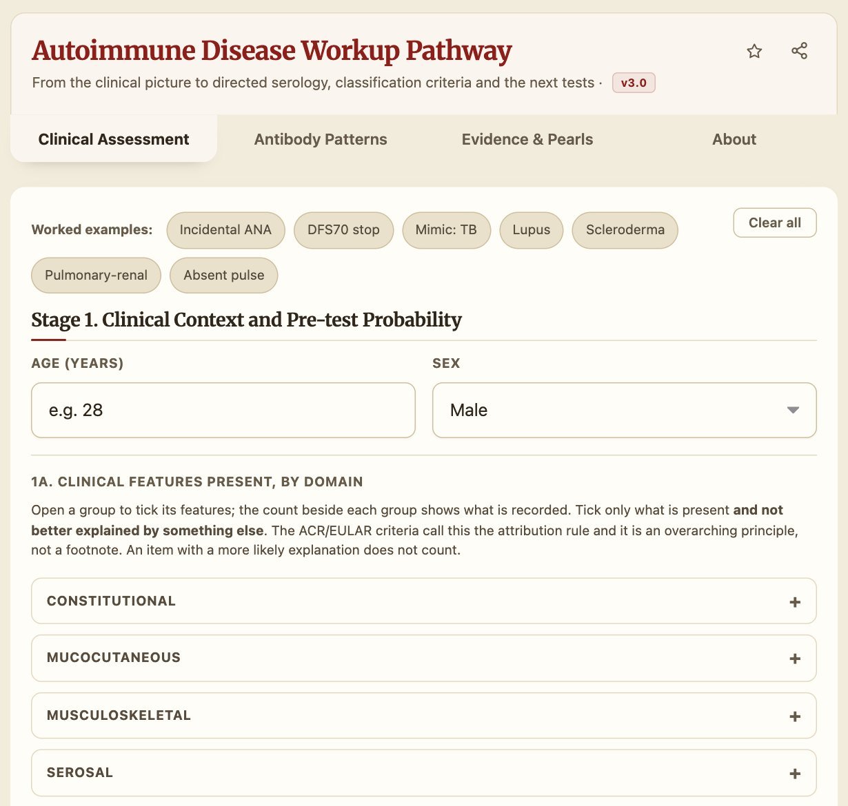
Stage 1. Clinical context
Enter the age and sex. Then open each group of features (constitutional, mucocutaneous, musculoskeletal and so on) and tick only what is present and not better explained by something else. This rule matters: the classification criteria count an item only when no more likely cause exists. The count beside each group shows how many you have ticked.
Two groups deserve a special mention. Features of diseases with no useful antibody lets you record signs of Takayasu arteritis, giant cell arteritis, polymyalgia, Behçet disease, spondyloarthritis and adult-onset Still disease. Organ-specific autoimmunity covers nephrotic syndrome, blistering skin disease, acute flaccid weakness, encephalitis and liver disease. Neither group is scored, but each produces a signpost to the test that confirms it.
If an infection or another condition could explain the picture (tuberculosis, endocarditis, HIV, hepatitis, a drug), tick it in the mimics list. The tool will then suspend classification until you have dealt with it.
The tool sets a pre-test probability from what you have entered. You can override it if your own judgement differs.
Stages 2 and 3. The ANA and the directed panel
Enter the ANA method, titre and pattern. The pattern decides which antibody groups Stage 3 opens, so the tool steers you towards the tests that fit. Then record each antibody as the report gives it: negative, weak positive, strong positive, or not done. A panel that was never sent is not a negative panel, and the tool keeps the two apart.
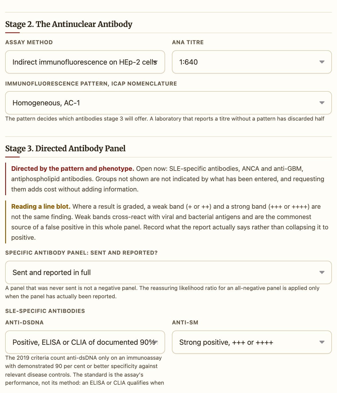
Stage 4. Confirmatory and organ-staging tests
Complement, urine findings, the renal biopsy class, lung imaging, sinus imaging and similar results go here. They complete the criteria and decide which organ alerts appear.
Reading the result
The result opens with an At a Glance card, a one-line summary of what was entered, and the classification verdict.
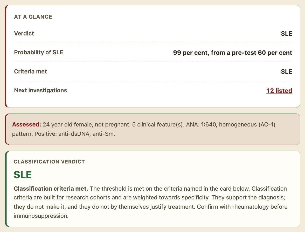
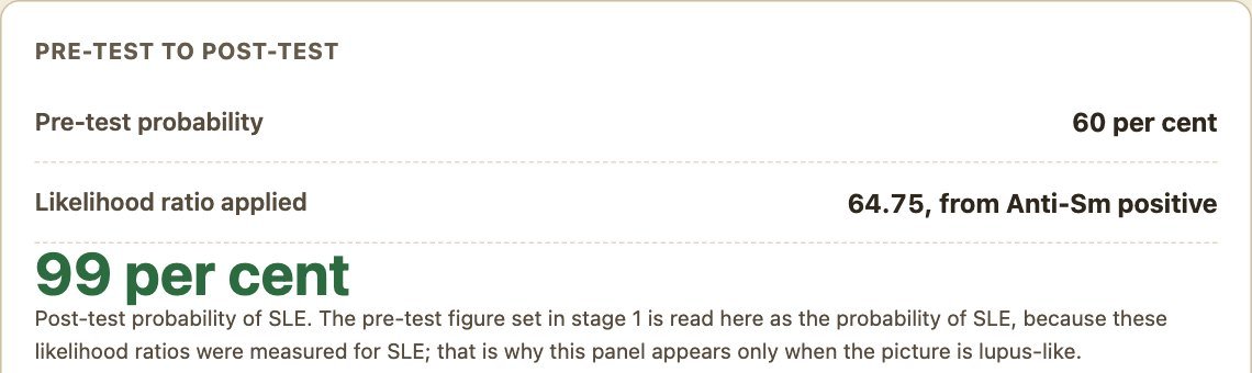
When the picture is lupus-like, the tool shows a pre-test to post-test panel. It applies only the single strongest likelihood ratio present and names it. Autoantibodies travel together, so multiplying their likelihood ratios one after another would overstate the result. The likelihood ratios come from a study of patients referred for testing in one Chinese hospital [15], so read the figure as an order of magnitude, not a measurement. When the picture is not lupus-like, for example arthritis with rheumatoid serology, the tool withholds the figure rather than give a probability of the wrong disease.
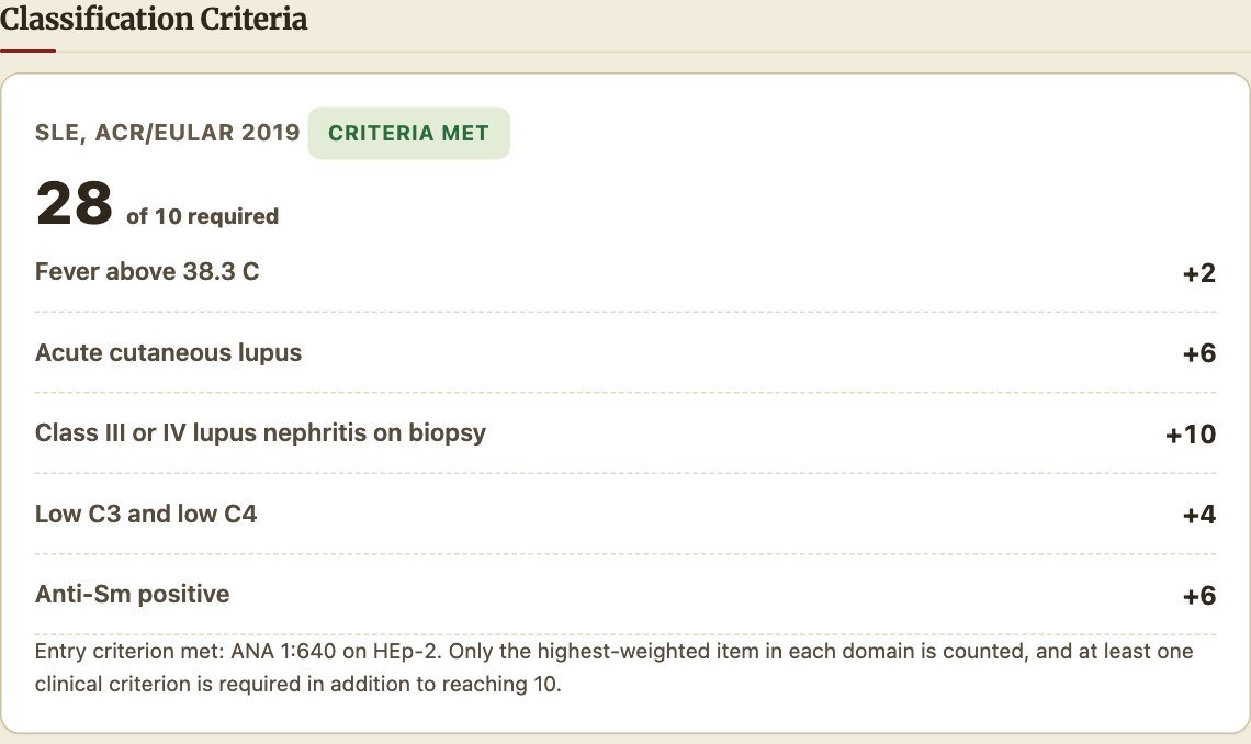
Each set of classification criteria gets a card that lists every item counted and its weight, so you can check the arithmetic yourself.
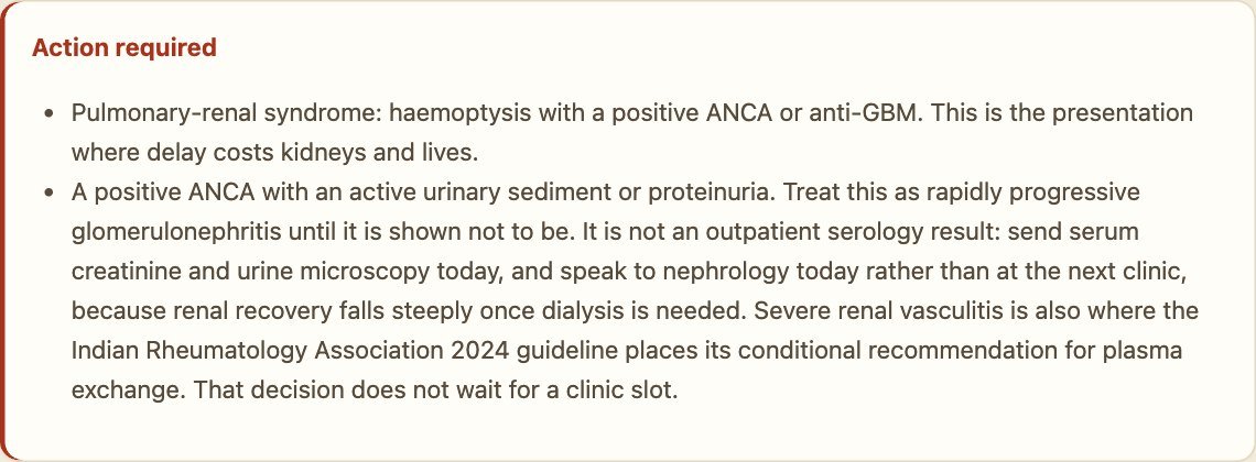
If anything needs same-day action, an Action required box appears above the scores. A positive ANCA with haemoptysis or an active urine sediment is one example.
Below these come the antibody interpretation, the signposts for conditions the antibody panel cannot answer, and a list of next investigations. The list is built from what you entered and ends with the screen to complete before immunosuppression. Nothing in it changes a score or the verdict.
Saving, printing and sharing
At the bottom of the page you can copy the result for the electronic record, print it as a PDF (with or without references), or use Share this calculation with a colleague. The share link carries the entered findings, and no name, so a colleague who opens it sees exactly what you saw. The worked cases below are share links of this kind.
How to use the Autoantibody Interpretation Guide
Open the Autoantibody Interpretation Guide when you have an antibody report and want to know what it means. It does not ask about symptoms, and it gives no score or probability.
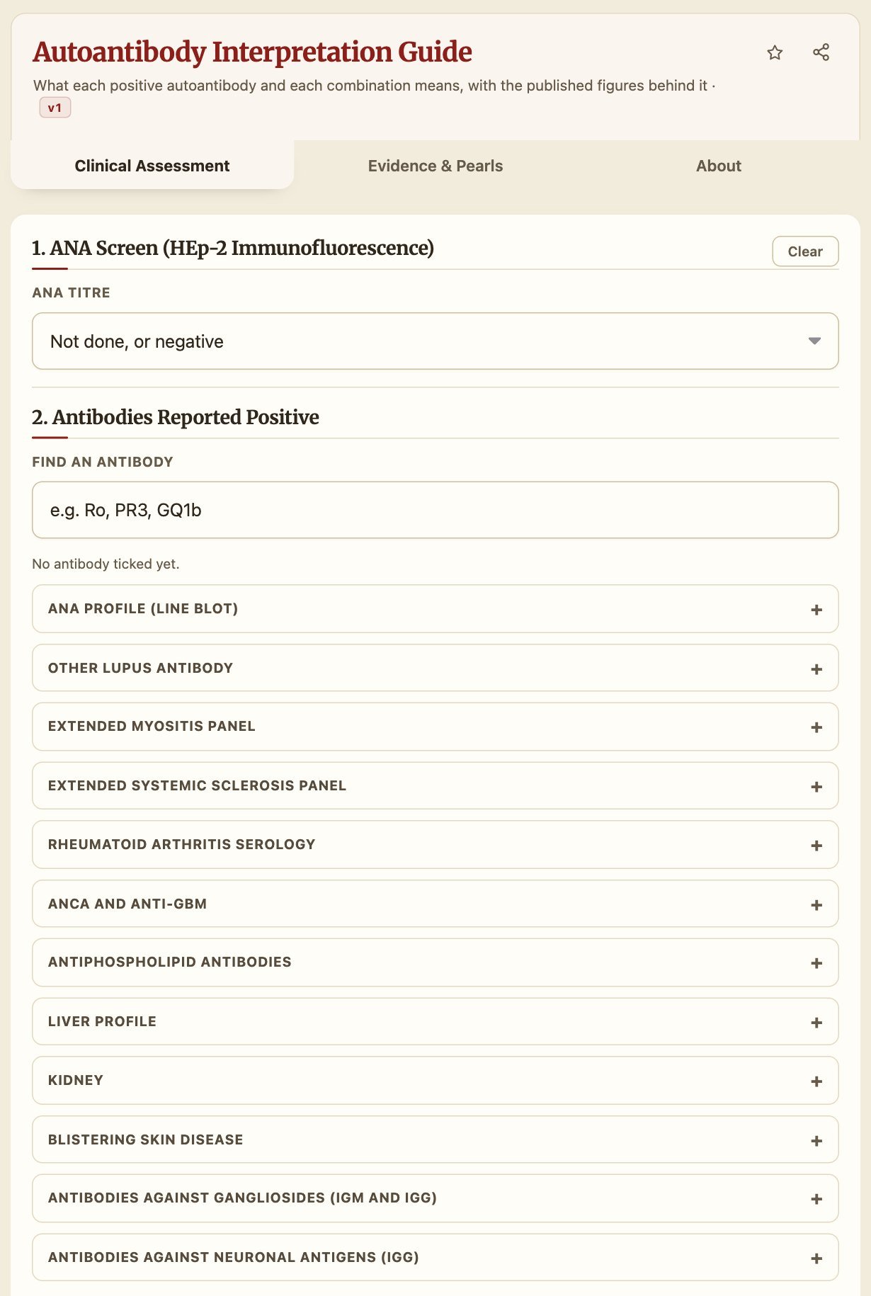
- Enter the ANA titre and pattern, if an ANA was done.
- Tick each antibody the report shows as positive. Type part of a name (for example Ro, PR3 or GQ1b) in Find an antibody, or open the panel it came from. The panels follow the way laboratories group their tests: ANA profile, myositis, systemic sclerosis, rheumatoid arthritis, ANCA and anti-GBM, antiphospholipid, liver, kidney, blistering skin disease, gangliosides and neuronal antigens.
- Where a panel asks for more detail, add it. For antiphospholipid antibodies, that is the IgG level and whether the result was positive again 12 weeks later. For neuronal antibodies, it is how the result was confirmed.
- Read the result.
Leaving an antibody unticked is not the same as recording it negative. The form cannot tell “not tested” from “negative”, so any conclusion that depends on a negative test is written as “if it was tested and negative”.
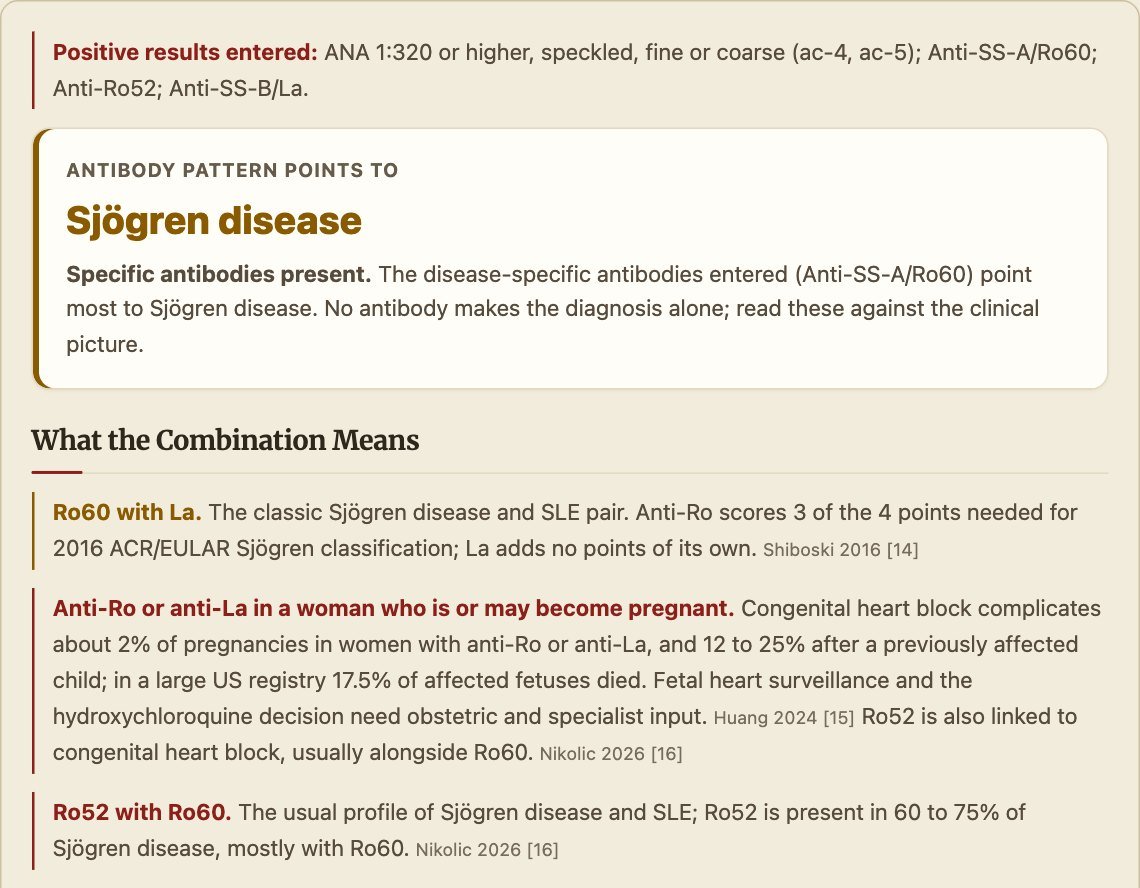
The result starts with the pattern the antibodies point to, then explains what the combination means, with its source beside each statement.
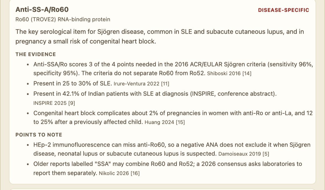
Each positive antibody then gets its own card: how specific it is, the published figures behind it, and the points to note.
The guide also carries a short explanation for the patient and family, and a link to the workup tool for when you need the clinical picture and the criteria.
Worked cases in the workup tool
Each link opens the tool with the case already entered. All the cases are fictional. Try changing one finding, for example removing the biopsy in case 1 or the endocarditis flag in case 4, and see what changes.
Case 1. Lupus nephritis (classical)
A 24-year-old woman with fever, a malar rash, mouth ulcers, proteinuria and an active urine sediment. ANA 1:640 homogeneous; anti-dsDNA positive on an assay of documented 90 per cent specificity; anti-Sm strongly positive; low C3 and C4; renal biopsy class III or IV lupus nephritis.
What the tool shows: SLE, 28 points against the 10 required; probability of SLE 99 per cent from a pre-test 60 per cent; 12 next investigations, including the infection screen before immunosuppression.
What to notice: the mouth ulcers and the proteinuria do not add points, because only the highest item in each domain counts (the malar rash and the biopsy). Anti-dsDNA and anti-Sm share a domain, so only one of them scores. Open case 1
Case 2. Rheumatoid arthritis (classical)
A 48-year-old woman with more than 10 swollen joints including small joints, for more than six weeks. RF and anti-CCP both high positive; raised CRP; ANA 1:80 speckled.
What the tool shows: rheumatoid arthritis, 10 points against the 6 required. The lupus probability is withheld.
What to notice: a low-titre ANA is common in rheumatoid arthritis and in healthy people. Joint involvement alone gives 6 lupus points, but the lupus criteria are not met, and the tool refuses to calculate a probability of lupus for what is clearly a rheumatoid picture. Open case 2
Case 3. Granulomatosis with polyangiitis (classical)
A 52-year-old man with nasal crusting and bleeding, hearing loss, haemoptysis, proteinuria and an active sediment. ANA negative; PR3-ANCA positive; lung nodules; sinus inflammation on imaging; pauci-immune glomerulonephritis on biopsy.
What the tool shows: GPA, 13 points against the 5 required, and an Action required box for a pulmonary-renal syndrome.
What to notice: the alert comes before the score. This patient needs a creatinine, urine microscopy and a nephrologist today, not at the next clinic. The negative ANA means the lupus criteria cannot be applied at all. Open case 3
Case 4. MPO-ANCA, low complement and haematuria: vasculitis or endocarditis? (confusing)
A 45-year-old man with fever, arthritis and an active urine sediment. ANA 1:80 speckled, MPO-ANCA positive by ELISA, one complement component low. Infective endocarditis is flagged as a possible explanation.
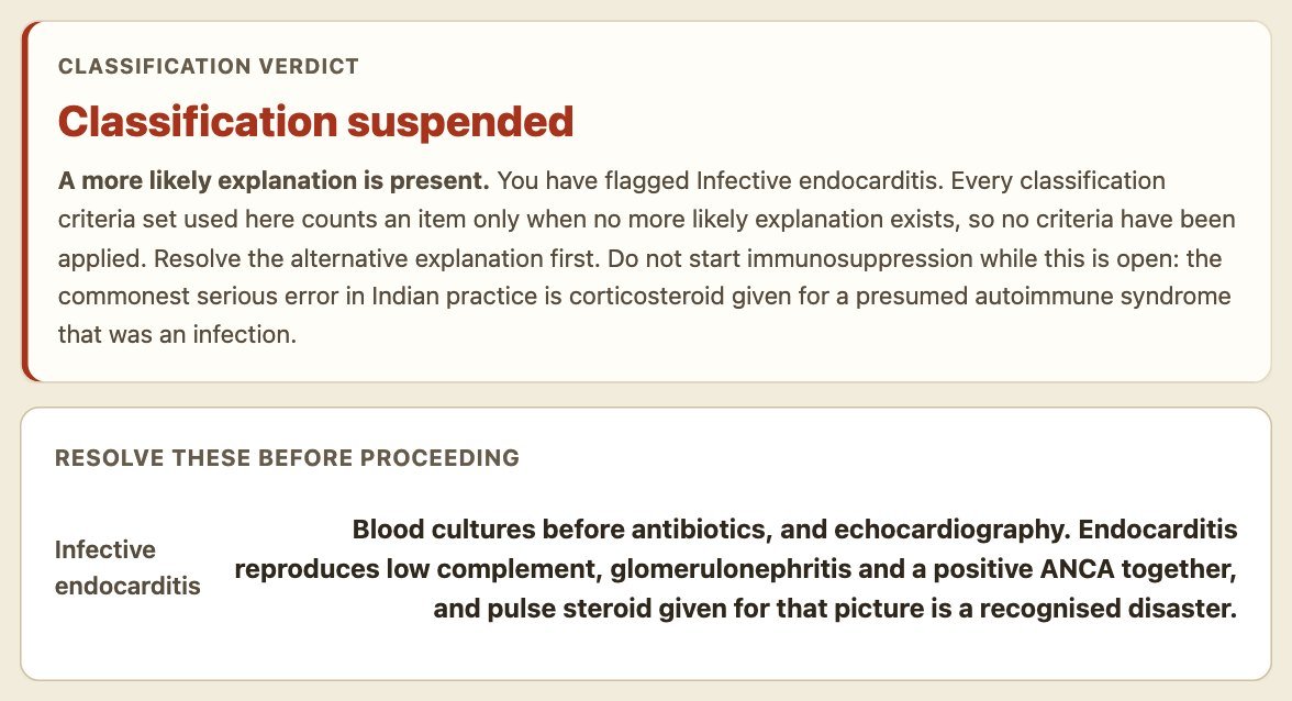
What the tool shows: classification suspended. No criteria are applied while the endocarditis question is open.
What to notice: endocarditis can produce fever, glomerulonephritis, low complement and a positive ANCA all at once, and MPO-ANCA is the least specific result in the panel. Pulse steroids for this picture, given to a patient with endocarditis, can kill. Take the blood cultures and do the echocardiogram first. Open case 4
Case 5. Fever, arthritis, rash and a low-titre ANA: lupus or adult-onset Still disease? (confusing)
A 23-year-old woman with daily spiking fever, a salmon-pink rash that comes and goes with the fever, and arthritis. ANA 1:80 speckled; RF and anti-CCP negative; raised CRP.
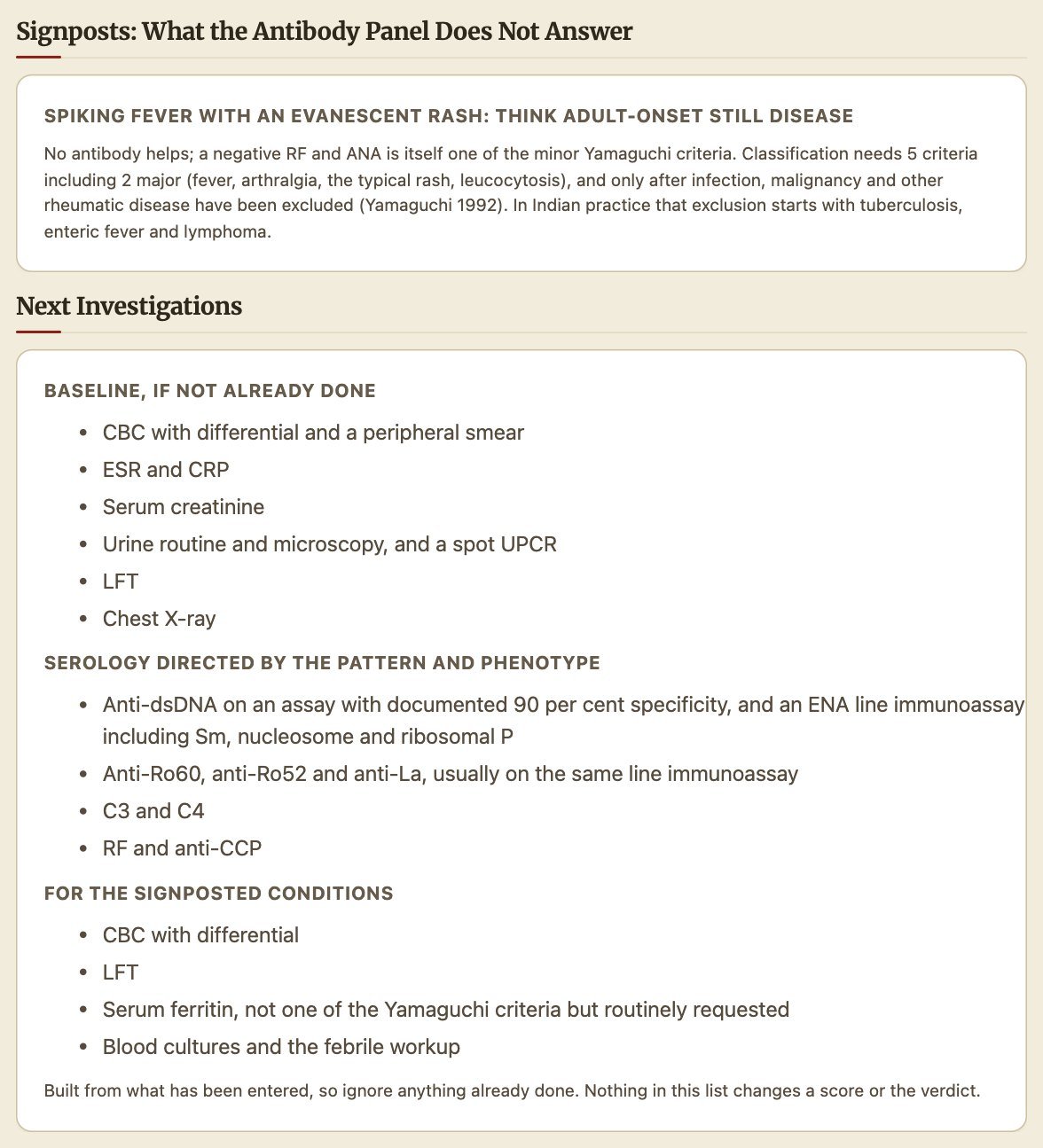
What the tool shows: SLE 8 points against the 10 required, not met; no post-test probability; a signpost to adult-onset Still disease.
What to notice: 8 points can tempt you to call it early lupus. But a 1:80 ANA is found in about one healthy person in eight, and nothing here is lupus-specific. The fever pattern and rash point to Still disease, which has no antibody and is a diagnosis of exclusion. In India that exclusion starts with tuberculosis, enteric fever and lymphoma. Open case 5
The tool also has seven built-in examples (Incidental ANA, DFS70 stop, Mimic: TB, Lupus, Scleroderma, Pulmonary-renal, Absent pulse). Tap any of them at the top of the blank form.
Worked cases in the antibody guide
Case 6. Anti-Ro60, anti-Ro52 and anti-La
ANA 1:320 speckled with Ro60, Ro52 and La positive. The guide points to Sjögren disease, notes that anti-Ro gives 3 of the 4 points needed for the 2016 Sjögren criteria [11], and warns about congenital heart block if the patient is or may become pregnant. Open case 6
Case 7. Anti-Jo-1 with anti-Ro52 and a cytoplasmic ANA
ANA 1:160 cytoplasmic, anti-Jo-1 and anti-Ro52 positive. The guide points to antisynthetase syndrome and explains why Ro52 with Jo-1 raises the concern about lung disease. It also reminds you that a cytoplasmic pattern can hide behind an “ANA negative” report. Open case 7
Case 8. Triple-positive antiphospholipid antibodies
Lupus anticoagulant, anticardiolipin IgG and anti-beta2-glycoprotein I IgG, at a high level and positive again 12 weeks later. The guide points to antiphospholipid syndrome and explains why this is the highest-risk profile.
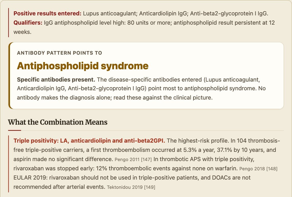
In thrombosis-free triple-positive carriers, a first clot occurred at 5.3 per cent a year [16]. In triple-positive patients with thrombosis, rivaroxaban did worse than warfarin [17], and EULAR advises against rivaroxaban in this group [18]. Remember the 12-week rule: a single positive result, often after an infection, is not antiphospholipid syndrome. Open case 8
Case 9. Isolated anti-DFS70
ANA 1:320 dense fine speckled, with anti-DFS70 the only antibody. The guide calls this a pattern usually seen in healthy people, provided no disease-specific antibody was found and the clinical picture does not suggest a rheumatic disease.
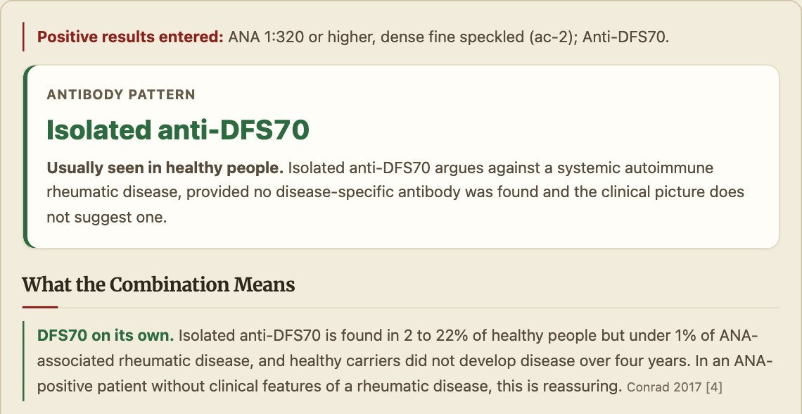
This is the result that lets you reassure a well person with a high-titre ANA, and stop testing. Open case 9
Ten common mistakes, and how to avoid them
- Ordering an ANA with no clinical question. Decide first what you would do with each result.
- Calling a low-titre ANA “lupus”. At 1:80, about one healthy person in eight is positive.
- Accepting an ANA report with no pattern. Ask the laboratory for it.
- Missing a cytoplasmic pattern reported as “negative” in a patient with myositis or lung disease.
- Ordering a large panel for everyone instead of the antibodies the pattern and the picture suggest.
- Treating a positive MPO-ANCA on its own. Look for a vasculitic syndrome, and exclude endocarditis, tuberculosis and drugs.
- Diagnosing antiphospholipid syndrome on a single result. Repeat it after at least 12 weeks.
- Repeating static markers such as the ANA, anti-Sm or anti-Ro. They do not change with activity.
- Stopping at a negative panel. Takayasu arteritis, Still disease, Behçet disease and spondyloarthritis have no antibody.
- Starting steroids before the infection screen. Tuberculosis, hepatitis B and HIV come first.
What the tools do not do
Both tools support clinical judgement; they do not replace it, and they do not replace a rheumatologist. The workup tool does not cover children, does not calculate disease activity scores such as the SLEDAI, and applies the 2006 Sydney criteria for antiphospholipid syndrome rather than the more restrictive 2023 criteria. The antibody guide computes no probability and no score. The list of what each tool does and does not cover is on its About tab, with every reference in full.
The workup tool was reviewed by the author on 24 September 2026, and an independent review by a rheumatologist is to follow. The antibody guide was reviewed by the author on 28 September 2026.
Declaration: the author built and maintains both tools. They are free to use.
References
- Satoh M, Chan EKL, Ho LA, et al. Prevalence and sociodemographic correlates of antinuclear antibodies in the United States. Arthritis Rheum. 2012;64(7):2319-2327.
- Tan EM, Feltkamp TE, Smolen JS, et al. Range of antinuclear antibodies in “healthy” individuals. Arthritis Rheum. 1997;40(9):1601-1611.
- Leuchten N, Hoyer A, Brinks R, et al. Performance of antinuclear antibodies for classifying systemic lupus erythematosus: a systematic literature review and meta-regression of diagnostic data. Arthritis Care Res (Hoboken). 2018;70(3):428-438.
- Damoiseaux J, Andrade LEC, Carballo OG, et al. Clinical relevance of HEp-2 indirect immunofluorescent patterns: the International Consensus on ANA patterns (ICAP) perspective. Ann Rheum Dis. 2019;78(7):879-889.
- Conrad K, Röber N, Andrade LE, Mahler M. The clinical relevance of anti-DFS70 autoantibodies. Clin Rev Allergy Immunol. 2017;52(2):202-216.
- Bossuyt X, Cohen Tervaert JW, Arimura Y, et al. Position paper: revised 2017 international consensus on testing of ANCAs in granulomatosis with polyangiitis and microscopic polyangiitis. Nat Rev Rheumatol. 2017;13(11):683-692.
- Miyakis S, Lockshin MD, Atsumi T, et al. International consensus statement on an update of the classification criteria for definite antiphospholipid syndrome (APS). J Thromb Haemost. 2006;4(2):295-306.
- Mahr A, Batteux F, Tubiana S, et al. Brief report: prevalence of antineutrophil cytoplasmic antibodies in infective endocarditis. Arthritis Rheumatol. 2014;66(6):1672-1677.
- Aringer M, Costenbader K, Daikh D, et al. 2019 European League Against Rheumatism/American College of Rheumatology classification criteria for systemic lupus erythematosus. Arthritis Rheumatol. 2019;71(9):1400-1412.
- Aletaha D, Neogi T, Silman AJ, et al. 2010 rheumatoid arthritis classification criteria: an American College of Rheumatology/European League Against Rheumatism collaborative initiative. Arthritis Rheum. 2010;62(9):2569-2581.
- Shiboski CH, Shiboski SC, Seror R, et al. 2016 American College of Rheumatology/European League Against Rheumatism classification criteria for primary Sjögren’s syndrome. Ann Rheum Dis. 2017;76(1):9-16.
- Robson JC, Grayson PC, Ponte C, et al. 2022 American College of Rheumatology/European Alliance of Associations for Rheumatology classification criteria for granulomatosis with polyangiitis. Ann Rheum Dis. 2022;81(3):315-320.
- Yamaguchi M, Ohta A, Tsunematsu T, et al. Preliminary criteria for classification of adult Still’s disease. J Rheumatol. 1992;19(3):424-430.
- Fragoulis GE, Nikiphorou E, Dey M, et al. 2022 EULAR recommendations for screening and prophylaxis of chronic and opportunistic infections in adults with autoimmune inflammatory rheumatic diseases. Ann Rheum Dis. 2023;82(6):742-753.
- Li H, Zheng Y, Chen L, Lin S. High titers of antinuclear antibody and the presence of multiple autoantibodies are highly suggestive of systemic lupus erythematosus. Sci Rep. 2022;12(1):1687.
- Pengo V, Ruffatti A, Legnani C, et al. Incidence of a first thromboembolic event in asymptomatic carriers of high-risk antiphospholipid antibody profile: a multicenter prospective study. Blood. 2011;118(17):4714-4718.
- Pengo V, Denas G, Zoppellaro G, et al. Rivaroxaban vs warfarin in high-risk patients with antiphospholipid syndrome. Blood. 2018;132(13):1365-1371.
- Tektonidou MG, Andreoli L, Limper M, et al. EULAR recommendations for the management of antiphospholipid syndrome in adults. Ann Rheum Dis. 2019;78(10):1296-1304.