

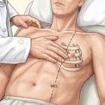





It is late evening on the medical ward. A 58-year-old woman, a cashew factory worker from Karkala, has had watery diarrhoea for four days. She takes hydrochlorothiazide for her blood pressure. She is weak, and her legs feel heavy. Her potassium is 2.6 mEq/L. The ECG shows prominent U waves. The intern has already written for intravenous potassium chloride in dextrose saline. This is an illustrative case, not a real patient.
“How much potassium can I give, and how fast?” By the end of this article you will know the limits, why the potassium sometimes refuses to rise however much you give, and how to find out why it fell.
Use this at the bedside
Open the tool: Acute Hypokalaemia
Get the CDSS app: install the MEDiscuss CDSS on your phone
1. Check the Magnesium First
About 40 to 60 per cent of patients with a low potassium also have a low magnesium. And until the magnesium is corrected, the hypokalaemia is refractory to potassium replacement. Why?
Magnesium depletion activates ROMK (Renal Outer Medullary Potassium) channels in the distal nephron, so the kidney goes on wasting potassium however much KCl is given.
Think of filling a tank with the drain open. Potassium goes in through the drip and leaves through the kidney just as fast. Magnesium closes the drain. So if several doses of KCl have gone in and the potassium is not rising, the most likely cause is uncorrected hypomagnesaemia. Check the serum magnesium, and correct it, before escalating the KCl.
Our patient has lost both through her bowel, and her thiazide wastes both through her kidney. Her magnesium is sent with the next sample.
2. The Limits for Intravenous Potassium
Intravenous potassium chloride is a high-alert medication. An error in rate or concentration causes fatal hyperkalaemia and cardiac arrest. So the limits in the table are not suggestions: they are absolute.
| Parameter | Peripheral IV | Central Line | Cardiac Arrest |
|---|---|---|---|
| Maximum Rate | 10 mEq/hour | 20 mEq/hour | 40 mEq/hour (ICU only) |
| Maximum Concentration | 40 mEq/L | 60 to 80 mEq/L | As per protocol |
| Preferred Diluent | Normal Saline (0.9% NaCl). Never use dextrose-containing fluids - insulin release from dextrose drives K+ intracellularly and worsens hypokalaemia. | ||
| Monitoring | Repeat K+ every 4 h | Continuous telemetry + K+ every 2 h | Continuous telemetry + K+ every 1 h |
“Why not dextrose? She is not eating.” Look at the diluent row. Dextrose releases insulin, and insulin drives potassium into the cells, so the serum potassium falls further while you think you are replacing it. Our intern’s dextrose saline is changed to normal saline.
Never give undiluted KCl as a bolus push: it causes instant local hyperkalaemia in the conduction system, and ventricular fibrillation within seconds. And through a peripheral vein, concentrations above 40 mEq/L cause severe phlebitis and pain. If the patient reports burning at the drip site, slow the rate or dilute further.
3. By Mouth Whenever Possible
Oral replacement is safer, more physiological and preferred whenever the patient can swallow and absorb it. Absorption is efficient, and the risk of overshoot is lower than with the intravenous route.
| Preparation | Brands | K+ Content | Notes |
|---|---|---|---|
| KCl syrup (elixir) | Potklor | 20 mEq per 15 mL | Bitter taste. Mix with juice. Commonest form in Indian hospitals. |
| KCl slow-release tablets | Check local stock | 8 mEq (600 mg) per tablet | Better tolerated. Do not crush, which defeats the slow-release coat. We could not verify which slow-release brands are stocked in India, so none is named here. |
| Potassium Citrate | Check local stock | Variable | Preferred in RTA and metabolic acidosis, because it provides alkali. Not ideal for routine replacement. We could not verify which potassium citrate brands are stocked in India, so none is named here. |
The main limit is the gut: nausea, vomiting, cramps and diarrhoea, all dose-related. Dividing the dose across the day, 20 mEq three times daily with meals, reduces intolerance significantly.
4. Reading the ECG
Our patient’s ECG had prominent U waves. The table places that finding.
| Stage | ECG Finding | Typical K+ Range | Clinical Significance |
|---|---|---|---|
| Early | ST segment depression, T wave flattening | 3.0 - 3.5 mEq/L | Subtle and easily missed. Compare with prior ECGs. |
| Moderate | Prominent U waves (best seen in V2-V3), apparent QT prolongation (actually QU prolongation) | 2.5 - 3.0 mEq/L | U wave is the hallmark ECG finding of hypokalaemia. May be confused with a long QT. |
| Severe | T-U wave fusion, ST depression deepens, PR prolongation | 2.0 - 2.5 mEq/L | Increased risk of atrial and ventricular ectopy. |
| Life-threatening | VT, VF, Torsades de Pointes, asystole | < 2.0 mEq/L | Cardiac arrest. Often triggered by concurrent hypomagnesaemia or digoxin. |
A common mistake: calling it a long QT and reaching for the QT drug list. The U wave is the hallmark of hypokalaemia, and when it merges with the T wave the interval you are measuring is really a QU interval. The other mistake is to trust the table’s ranges too much. The changes may not track the serum potassium closely, and some patients develop arrhythmias at mild levels, especially on digoxin or with a low magnesium.
Digoxin deserves its own line. Digoxin and potassium compete for the same binding site on the Na+/K+-ATPase pump, so as the potassium falls more digoxin binds, and a normal digoxin level does not exclude toxicity. In a patient on digoxin, keep the potassium above 4.0 mEq/L at all times, with a target of 4.0 to 5.0.
5. The Deficit Is Larger Than It Looks
Serum potassium is only 2 per cent of the total body potassium. The rest is inside the cells. So the serum level always underestimates how much has been lost. At a serum potassium of 2.5 to 2.9 mEq/L, the deficit is roughly 200 to 400 mEq, and full correction takes 2 to 3 days, often with oral and intravenous potassium together.
“So should I give 400 mEq today?” No. The first goal is a safe level, above 3.0 mEq/L, reached quickly. The rest of the replacement follows over 2 to 5 days. The rate limits exist for a reason.
6. Why Did the Potassium Fall?
Every low potassium has a cause, and the causes fall into four groups: potassium moving into cells, loss through the kidney, loss through the gut, and too little going in.
| Transcellular Shift | Renal Losses | GI Losses | Inadequate Intake |
|---|---|---|---|
| Insulin therapy / DKA treatment | Loop diuretics (Furosemide) | Diarrhoea (most common GI cause) | Alcoholism / malnutrition |
| Beta-2 agonists (Salbutamol) | Thiazide diuretics (HCTZ) | Vomiting / NG suction (causes renal loss via metabolic alkalosis) | Anorexia nervosa |
| Metabolic alkalosis | Hyperaldosteronism (Conn syndrome) | Laxative abuse | Tea-and-toast diet (elderly) |
| Thyrotoxic periodic paralysis | Cushing syndrome / exogenous steroids | Villous adenoma of colon | Prolonged NPO without K supplementation |
| Hypothermia / refeeding syndrome | RTA Type 1 and Type 2 | Fistulae / ostomy output | |
| Bartter / Gitelman syndrome | |||
| Amphotericin B / Cisplatin nephrotoxicity |
“How do I tell the kidney from the gut?” Send a spot urine potassium. In the setting of hypokalaemia, a urine potassium above 30 mEq/L suggests renal wasting. A transtubular potassium gradient (TTKG) above 4 suggests inappropriate renal potassium secretion, as with aldosterone excess or diuretics. Our patient may well have both: the diarrhoea from her gut, and the thiazide working on her kidney.
7. The Patient With Diabetic Ketoacidosis
One group needs a warning of its own. In diabetic ketoacidosis the total body potassium is always depleted, typically by 200 to 600 mEq, even when the serum potassium on arrival is normal or high. Start insulin, and the potassium moves back into the cells within minutes. So the rule from the 2024 ADA, EASD and JBDS consensus is firm: do not start insulin if the serum potassium is below 3.5 mmol/L. Replace potassium first, at 10 to 20 mmol/h of intravenous KCl, and start the insulin once it is above 3.5.
Back to Our Patient
Her drip is changed to normal saline, with KCl at no more than 10 mEq/hour and 40 mEq/L through her peripheral line, and her potassium is repeated every 4 hours. Her magnesium comes back low and is corrected. Once she can take it, she is given KCl syrup in divided doses. The thiazide is held. Her deficit is probably 200 to 400 mEq, so the team plans 2 to 3 days of replacement rather than one heroic night.
Try This
A 24-year-old man with type 1 diabetes arrives in diabetic ketoacidosis. His potassium is 3.2 mmol/L. The resident wants to start the insulin infusion now and add potassium to the fluids. What would you do? Illustrative case.
Answer. Hold the insulin. His potassium is below 3.5 mmol/L, and insulin would drive it lower still. Replace potassium first, at 10 to 20 mmol/h of intravenous KCl, and start the insulin once it is above 3.5. Above 10 mmol/h, a central line and telemetry are required.
Take-Home Points
- If the potassium will not rise, check and correct the magnesium before giving more KCl.
- Peripherally, no more than 10 mEq/hour and 40 mEq/L; never a bolus; never in dextrose.
- Give it by mouth whenever possible, in divided doses with meals.
- The serum level underestimates the deficit: aim for above 3.0 quickly, then replace over days.
- In diabetic ketoacidosis, do not start insulin with a potassium below 3.5 mmol/L.
For the opposite problem, see the queue’s article Acute Hyperkalaemia: The Evidence Behind the Protocol. For what potassium and magnesium do to the QT interval, see The QT Interval and the Electrolytes: The Evidence Behind Two Tools; for the potassium that falls when a starved patient is fed, Refeeding Syndrome: The Evidence Behind the Pathway.
Use this at the bedside
Open the tool: Acute Hypokalaemia
Get the CDSS app: install the MEDiscuss CDSS on your phone
Declaration: the author built and maintains the tool. It is free to use. The figures in this article are those the tool carried in its Evidence tab on 5 November 2026, each from the source the tool names. The illustrative cases follow the tool’s rules; check the tool’s result for a real patient.
References
- Unwin RJ, Luft FC, Shirley DG. Pathophysiology and management of hypokalemia: a clinical perspective. Nat Rev Nephrol. 2011;7(2):75-84.
- Crop MJ, Hoorn EJ, Lindemans J, Zietse R. Hypokalaemia and subsequent hyperkalaemia in hospitalized patients. Nephrol Dial Transplant. 2007;22(12):3471-3477.
- Huang CL, Kuo E. Mechanism of hypokalemia in magnesium deficiency. J Am Soc Nephrol. 2007;18(10):2649-2652.
- Kardalas E, Paschou SA, Anagnostis P, et al. Hypokalemia: a clinical update. Endocr Connect. 2018;7(4):R135-R146.
- Umpierrez GE, Davis GM, ElSayed NA, et al. Hyperglycemic Crises in Adults With Diabetes: A Consensus Report. Diabetes Care. 2024;47(8):1257-1275.
- Indian Council of Medical Research (ICMR). Standard Treatment Workflows - Electrolyte Emergencies. 2019.
- Viera AJ, Wouk N. Potassium Disorders: Hypokalemia and Hyperkalemia. Am Fam Physician. 2015;92(6):487-495.
- Palmer BF, Clegg DJ. Physiology and Pathophysiology of Potassium Homeostasis: Core Curriculum 2019. Am J Kidney Dis. 2019;74(5):682-695.


Leave a Reply