

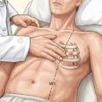





It is ten at night. A 66-year-old retired bank clerk from Hebri, with diabetes and chronic kidney disease, came in weak and breathless. He takes ramipril and spironolactone for his heart, and four days ago a general practitioner started cotrimoxazole for a urinary infection. His potassium has come back at 7.1 mEq/L. The ECG shows tall, peaked T waves and a long PR interval. This is an illustrative case, not a real patient.
“Do I give calcium first, or something to bring the potassium down?” By the end of this article you will know why calcium goes first, what each of the other treatments does and how quickly, and how to find out why his potassium rose.
Use this at the bedside
Open the tool: Acute Hyperkalaemia
Get the CDSS app: install the MEDiscuss CDSS on your phone
1. Why calcium goes first
Calcium does not lower serum potassium. So why is it the first drug? Because the danger of a high potassium is not the number. It is what the potassium does to the heart muscle’s electrical membrane.
It antagonises the membrane effects of hyperkalaemia. A high potassium raises the resting membrane potential, making it less negative and bringing it closer to the threshold potential, which causes initial hyper-excitability and then inexcitability: conduction blocks, asystole. Calcium raises the threshold potential and restores the normal voltage gap.
Put simply: every heart cell sits at a resting voltage, some distance from the voltage at which it fires. A high potassium closes that distance, so the cell first fires too easily and then cannot fire at all. Calcium moves the firing point back. The potassium is still high; the heart is temporarily protected. That buys time for the treatments that move the potassium.
The dose that is often wrong. Calcium Gluconate 10% carries 93 mg of elemental calcium, 2.3 mmol, in each 10 mL ampoule; Calcium Chloride 10% carries 272 mg, 6.8 mmol, in the same volume. So the gluconate dose is 30 mL and the chloride dose is 10 mL: both deliver 6.8 mmol. Ten millilitres of gluconate delivers 2.2 mmol and is an underdose, yet a great deal of teaching material still gives it. The UK Kidney Association guideline of December 2023 and a national patient safety alert of June 2023 both put the dose at 30 mL over 10 minutes. Gluconate goes peripherally; chloride goes through a central line only.
2. What each treatment does, and how fast
Only dialysis and the binders take potassium out of the body. Everything else either moves it into cells or protects the heart while the potassium is still there. The table puts each treatment beside how it works, how far it lowers the potassium, how soon it starts and how long it lasts. Read the last two columns most carefully: they tell you when the potassium will come back.
| Intervention | Mechanism | Expected K+ Drop | Onset | Duration |
|---|---|---|---|---|
| Calcium Gluconate 10% IV | Membrane stabilisation (no K+ change) | 0 mEq/L (protects heart only) | 1 - 3 min | 30 - 60 min |
| Insulin 10U + Dextrose 25g IV | Intracellular shift via Na+/K+-ATPase | 0.5 - 1.2 mEq/L | 15 - 30 min | 4 - 6 h |
| Salbutamol 10-20 mg nebulised | Intracellular shift via Beta-2 stimulation | 0.5 - 1.5 mEq/L | 15 - 30 min | 2 - 4 h |
| NaHCO3 (if acidotic) | Shift via pH correction | 0.3 - 0.5 mEq/L | 15 - 30 min | 1 - 2 h |
| Furosemide 40-80 mg IV | Renal elimination (kaliuresis) | Variable (requires renal function) | 15 - 30 min | 4 - 6 h |
| SZC (Lokelma) 10g PO | GI potassium binding | 0.4 - 0.7 mEq/L | 1 - 2 h | Ongoing |
| CPS / K-Bind 30g PO/PR | GI cation exchange | 0.5 - 1.0 mEq/L (over hours-days) | 2 - 6 h | Variable |
| Haemodialysis | Extracorporeal elimination | 1.0 - 2.0 mEq/L per hour | Immediate | Duration of session |
“So insulin is the strongest shift. Is that the end of it?” Not quite. Insulin-dextrose brings a delayed hypoglycaemia that peaks at 2 to 3 hours, and it is the commonest complication of treating hyperkalaemia. Check GRBS every 30 minutes for 2 hours, then hourly for a further 4 hours. Salbutamol, nebulised at 10 to 20 mg, adds to insulin, but about 20 to 40% of ESRD patients are resistant to it, so it cannot be the whole answer in a dialysis patient. Sodium bicarbonate works only in significant metabolic acidosis (pH below 7.2). And the resins take hours to days; they are elimination adjuncts, not emergency treatment.
Never run calcium gluconate and sodium bicarbonate through the same line without flushing. They precipitate into calcium carbonate crystals and block the line.
3. Reading the ECG
Our patient’s ECG had peaked T waves and a long PR interval. Where does that place him? The table lists the changes in the order they usually appear, with the potassium range at which each is typical and what it means.
| Stage | ECG Finding | Typical K+ Range | Clinical Significance |
|---|---|---|---|
| Early | Tall, peaked, narrow-based T waves (best seen in V2-V4) | 5.5 - 6.5 mEq/L | First ECG change. Often subtle. May be confused with hyperacute T waves of STEMI. |
| Moderate | PR prolongation, P wave flattening, ST depression | 6.5 - 7.5 mEq/L | Atrial conduction delay. P waves may become imperceptible. |
| Advanced | QRS widening (> 120 ms), bizarre QRS morphology | 7.0 - 8.0 mEq/L | Ventricular conduction delay. May mimic bundle branch block or ventricular tachycardia. |
| Pre-arrest | Sine wave pattern (fusion of widened QRS with T wave) | > 8.0 mEq/L | Imminent VF or asystole. This is a peri-arrest rhythm. Treat as cardiac arrest. |
A common mistake: treating the table as a ladder that every patient climbs one step at a time. The progression is not always sequential: some patients go directly from peaked T waves to cardiac arrest. Nor does it follow the serum level closely. A patient with a potassium of 7.0 may have a normal ECG, while one with 5.8 on digoxin may have advanced changes. Treat the ECG you see, and treat a sine wave as cardiac arrest.
4. Check the sample, then look for the cause
Before treating an unexpected high potassium in a stable patient, ask whether the sample is real. Was the tourniquet on too long? Was the sample haemolysed? Is there a very high white cell or platelet count? A fresh, atraumatic sample can prevent treatment the patient never needed. Our patient is unwell and his ECG has changed, so we treat first and repeat the sample alongside.
Then work through the causes. They fall into four groups: a false reading, potassium moving out of cells, the kidney failing to excrete it, and too much going in.
| Pseudohyperkalaemia | Transcellular Shift | Impaired Excretion | Increased Intake / Load |
|---|---|---|---|
| Prolonged tourniquet / fist clenching | Metabolic acidosis (DKA, lactic acidosis, RTA) | Acute Kidney Injury | Excessive IV KCl supplementation |
| In vitro haemolysis (traumatic draw) | Insulin deficiency / DKA | CKD Stage 4-5 / ESRD | Massive blood transfusion (stored blood) |
| Severe leukocytosis (> 70,000) | Non-selective beta-blockers | ACEi / ARBs / MRAs | Tumour lysis syndrome |
| Severe thrombocytosis (> 500,000) | Succinylcholine | NSAIDs / Calcineurin inhibitors | Rhabdomyolysis / crush injury |
| Delayed sample processing | Digoxin toxicity | TMP-SMX / Heparin | Major burns / haemolysis |
| Hypertonicity (mannitol, hyperglycaemia) | Hypoaldosteronism (Type 4 RTA) | High-K diet in CKD patients |
Our patient sits in the third column twice over: chronic kidney disease, and three drugs that impair excretion.
5. The drugs on his chart
Most hyperkalaemia on our wards has a drug somewhere in it. The table gives the common classes with the Indian brands you will see on the chart, how each raises the potassium, and what to do.
| Drug Class | Common Indian Brands | Mechanism | Action |
|---|---|---|---|
| ACE Inhibitors | Enalapril (Envas), Ramipril (Cardace, Ramistar) | Decreased aldosterone secretion | Hold / dose-reduce |
| ARBs | Losartan (Losar), Telmisartan (Telma, Telmikind) | Decreased aldosterone secretion | Hold / dose-reduce |
| MRAs | Spironolactone (Aldactone), Eplerenone (Epleheart) | Blocks aldosterone at collecting duct | Hold until K normalises |
| NSAIDs | Ibuprofen (Brufen), Diclofenac (Voveran) | Decreased renal blood flow and renin | Discontinue |
| TMP-SMX | Cotrimoxazole (Bactrim, Septran) | Blocks ENaC (amiloride-like effect) | Discontinue / substitute |
| Heparin | UFH, Enoxaparin (Clexane, Lonopin) | Suppresses aldosterone synthesis | Monitor; switch if possible |
| Potassium supplements | K-Lor, Potklor, IV KCl | Direct K+ load | Discontinue immediately |
“Cotrimoxazole? I thought it was just an antibiotic.” Its trimethoprim component blocks the epithelial sodium channel in the collecting duct, the same way the diuretic amiloride does, and the kidney cannot excrete potassium. In a man already on an ACE inhibitor and spironolactone with poor kidneys, four days of it is enough.
6. When dialysis is needed
Dialysis is the definitive elimination therapy; haemodialysis removes 25 to 50 mEq of potassium per hour. Call the nephrologist early for:
- an anuric or ESRD patient, in whom diuretics and renal elimination do not work;
- a potassium still at 6.0 or more after two full cycles of medical therapy;
- life-threatening ECG changes that do not respond to calcium gluconate;
- massive tissue breakdown, such as rhabdomyolysis, tumour lysis, major burns or crush injury;
- severe AKI with oliguria and a potassium rising despite shift therapies.
Back to our patient
He gets 30 mL of calcium gluconate 10% over 10 minutes, peripherally, and the ECG is repeated. Then insulin-dextrose (10 units of Actrapid with 25 g of dextrose) and nebulised salbutamol, with his GRBS checked every 30 minutes. Ramipril, spironolactone and cotrimoxazole are held and the infection treated with another drug. A fresh sample goes to the lab. The shift lasts hours, not days, so his potassium is rechecked; if it stays at 6.0 or more after two cycles, he needs dialysis.
Try this
A 40-year-old woman, well, with a routine potassium of 6.8 mEq/L. Her ECG is normal. Her platelet count is 720,000. The intern has drawn up calcium and insulin. What would you do first? Illustrative case.
Answer. Pause and check the sample. She is clinically stable with a normal ECG, and a platelet count above 500,000 is a cause of pseudohyperkalaemia: the potassium can rise in the tube, not in her. Repeat from a fresh, atraumatic draw before treating. A normal ECG does not by itself prove the potassium is false, which is why the repeat sample decides.
Take-home points
- Calcium protects the heart but does not lower the potassium: give 30 mL of gluconate 10%, not 10 mL.
- Insulin-dextrose and salbutamol move potassium into cells for hours; only dialysis and the binders remove it.
- Check the GRBS for 6 hours after insulin-dextrose: delayed hypoglycaemia is the commonest complication.
- The ECG does not track the potassium closely and need not climb step by step; treat the ECG you see.
- Look for the drug: ACE inhibitors, ARBs, MRAs, NSAIDs, cotrimoxazole, heparin and potassium supplements.
For what potassium, magnesium and calcium do to the QT interval, see the queue’s article The QT Interval and the Electrolytes: The Evidence Behind Two Tools. For the other common emergency electrolyte, see Acute Hyponatraemia: The Evidence Behind the Protocol.
Use this at the bedside
Open the tool: Acute Hyperkalaemia
Get the CDSS app: install the MEDiscuss CDSS on your phone
Declaration: the author built and maintains the tool. It is free to use. The figures in this article are those the tool carried in its Evidence tab on 5 November 2026, each from the source the tool names. The illustrative cases follow the tool’s rules; check the tool’s result for a real patient.
References
- Weisberg LS. Management of severe hyperkalemia. Crit Care Med. 2008;36(12):3246-3251.
- Rossignol P, Legrand M, Kosiborod M, et al. Emergency management of severe hyperkalemia: Guideline for best practice and opportunities for the future. Pharmacol Res. 2016;113(Pt A):585-591.
- Indian Council of Medical Research (ICMR). Standard Treatment Workflows - Acute Kidney Injury and Hyperkalaemia. 2019.
- Maxwell AP, Linden K, O’Donnell S, et al. Management of hyperkalaemia. J R Coll Physicians Edinb. 2013;43(3):246-251.
- Lindner G, Burdmann EA, Clase CM, et al. Acute hyperkalemia in the emergency department: a summary from a Kidney Disease: Improving Global Outcomes conference. Eur J Emerg Med. 2020;27(5):329-337.
- UK Kidney Association. Clinical Practice Guidelines: Treatment of Acute Hyperkalaemia in Adults. December 2023.
- Packham DK, Rasmussen HS, Lavin PT, et al. Sodium zirconium cyclosilicate in hyperkalemia. N Engl J Med. 2015;372(3):222-231.
- Batterink J, Cessford TA, Taylor RAI. Pharmacological interventions for the acute management of hyperkalaemia in adults. Cochrane Database Syst Rev. 2015;(10):CD010344.


Leave a Reply